

Background
Keratoacanthoma (KA) is a rapidly growing epithelial proliferation, clinically characterized by a nodular lesion with a central hyperkeratotic keratin plug, occurring predominantly on sun-exposed areas of elderly individuals. The clinical and histopathological similarities between KA and cutaneous squamous cell carcinoma (cSCC), particularly its well-differentiated variant, make definitive distinction difficult or even impossible in many cases; consequently, KA has historically been frequently reported as cSCC of the KA type.1 However, the well-recognized tendency of KA to regress spontaneously has led many authors to consider it a biologically benign tumor, with pathophysiological mechanisms distinct from those of malignant cSCC.2
Since its initial description, the therapeutic management of KA has remained controversial. Although a regressing KA may be monitored over several months, it is difficult to predict the maximum size reached before regression or how healing will occur. The diagnostic ambiguity between KA and cSCC has led to the general recommendation of surgical excision, following the same treatment standard applied to well-differentiated cSCC.3 While tumors located on the trunk and extremities can often be excised with relatively low esthetic and functional impact, patients with lesions in anatomically sensitive areas or with multiple KAs are more prone to greater surgical morbidity and disfigurement. Unfortunately, the skin of the head and neck is among the most frequently affected sites due to chronic sun exposure. The elderly are a particularly high-risk population who commonly present with facial lesions, multiple comorbidities, and limited physiological reserve.4 Several less invasive treatment modalities have been described, including cryotherapy, topical application of 5-fluorouracil (5-FU) or imiquimod, radiotherapy, photodynamic therapy, CO2 laser therapy, curettage with electrodessication, and intralesional (IL) therapies with bleomycin, 5-FU, interferon, and methotrexate (MTX).2,4,5
MTX-IL has emerged as a particularly attractive therapeutic option for KA. MTX is a folic acid analog that inhibits dihydrofolate reductase, thereby blocking nucleotide synthesis and interfering with cellular proliferation.6 The rationale for its IL use is based on the high proliferative rate of KA, with local administration maximizing therapeutic efficacy while minimizing systemic adverse effects.5,7 MTX-IL has been described both as a curative monotherapy and as a neoadjuvant approach, allowing for tumor volume reduction and facilitating subsequent surgical excision, with a positive impact on both esthetic and functional outcomes.7,8
Prospective and retrospective studies have reported complete response rates ranging from 70% to 100%, typically after two to six injections, with excellent local tolerability and minimal adverse effects.7,9,10 Comparative trials have demonstrated similar efficacy between MTX-IL and 5-FU, although 5-FU may achieve slightly faster lesion resolution at the expense of greater local inflammatory reaction.10,11
Given the scarcity of national data, the present work aims to describe the experience of a Portuguese dermatology department in the use of MTX-IL for the treatment of KA, thereby contributing to strengthening the available clinical evidence and supporting therapeutic decision-making in this condition.
Methods
We conducted a retrospective descriptive study of patients diagnosed with KA and treated with MTX-IL at our dermatology department. The study included all consecutive cases managed between 2024 and 2025.
Five cases of KA treated with MTX-IL were identified. Histopathological diagnosis was established by dermatopathologists according to standard criteria. Eligibility criteria were: (i) a solitary nodular KA suitable for IL therapy, (ii) availability of complete clinical records and follow-up, and (iii) absence of contraindications to MTX (immunosuppression, renal or hepatic impairment, pregnancy, or known hypersensitivity to MTX).
All procedures were performed in an outpatient clinical setting. MTX was administered intralesionally without anesthesia, as all patients demonstrated good procedural tolerance. A 30-gauge needle was used to inject MTX directly into the lesion. A standard dose of 25 mg (0.5 mL of a 50 mg/mL solution) was used in all cases, except for one patient with a smaller lesion, in whom a reduced dose of 12.5 mg (0.25 mL) was administered.
Injections were delivered at both the peripheral border and central portion of the nodular lesion, ensuring circumferential infiltration. As described in the previous series, partial leakage of the injected volume occurred consistently and was attributed to the low structural cohesion of KA tumor cells.
Treatments were repeated every 7 days until complete necrosis or clinically significant tumor reduction. The cumulative dose per lesion depended on the number of treatment sessions required. Baseline clinical assessment was performed in all patients, and none had known significant renal or hepatic impairment or immunosuppression. Given the low systemic exposure associated with IL administration and the small doses used, routine laboratory monitoring was not systematically performed.
Clinical response was assessed at each visit through measurement of maximum lesion diameter and evaluation of necrosis and regression. Complete response was defined as full clinical resolution without residual tumor. Adverse events were actively monitored at every session. Patients were followed for a minimum of 4 months after treatment completion to assess recurrence, residual scarring, or late adverse effects. Informed consent for publication of clinical images was obtained from all patients.
Results
A total of five patients with histologically confirmed KA were treated with MTX-IL (Table 1). The cohort included four men and one woman, with a mean age of 68.8 years (range: 63–77 years). Lesion diameter ranged from 5 mm to 20 mm, and lesions were located on cosmetically or functionally sensitive areas, including the nose (Fig. 1 and 2), preauricular region (Fig. 3), and hand (Fig. 4 and 5).
Table 1. Clinical characteristics and outcomes of patients treated with intralesional methotrexate for keratoacanthoma
| Patient | Age (years) | Sex | Lesion location | Lesion diameter | Duration before treatment | No. of injections | Clinical outcome | Follow-up | Recurrence |
|---|---|---|---|---|---|---|---|---|---|
| 1 | 71 | Male | Left nostril | 15 mm | 1 month | 7 | Complete resolution | 6 months | No |
| 2 | 63 | Female | Dorsum of the 2nd finger, left hand | 20 mm | 2 months | 3 | Complete resolution | 6 months | No |
| 3 | 64 | Male | Right preauricular area | 10 mm | 1.5 months | 2 | Complete resolution | 4 months | No |
| 4 | 71 | Male | Left nostril | 5 mm | 1 month | 1 | Complete resolution | 12 months | No |
| 5 | 77 | Male | Dorsum of the left hand | 10 mm | 3 months | 2 | Complete resolution | 8 months | No |
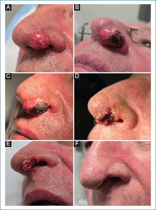
Figure 1. Clinical evolution of a keratoacanthoma of the left nostril treated with intralesional methotrexate. A: initial presentation showing a nodular keratotic lesion. B: one week after the first intralesional methotrexate injection, with evident inflammatory changes and early signs of tumor necrosis. C: after three injections, demonstrating increased necrosis and early tumor regression. D: after five injections, with marked reduction in lesion size. E: after six injections, showing continued involutive changes and further regression. F: final follow-up visit, with complete clinical resolution and an excellent cosmetic outcome.
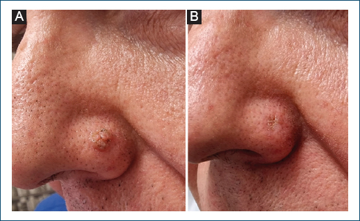
Figure 2. Clinical evolution of a keratoacanthoma of the left nostril treated with intralesional methotrexate. A: initial presentation showing a small nodular keratotic lesion. B: one month after a single intralesional methotrexate injection, demonstrating complete clinical resolution and a favorable cosmetic outcome.
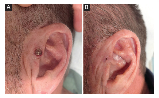
Figure 3. Clinical evolution of a keratoacanthoma of the right preauricular region treated with intralesional methotrexate. A: initial presentation showing a nodular lesion with a keratotic center. B: one week after the second and final intralesional methotrexate injection, with near-complete clinical resolution.
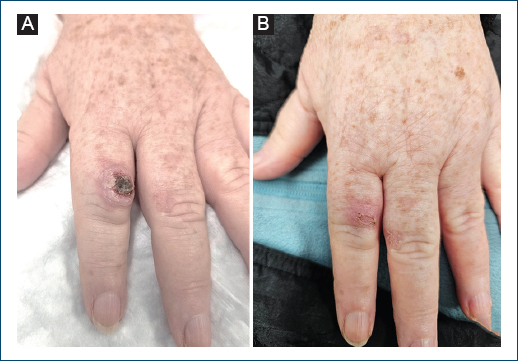
Figure 4. Clinical evolution of a keratoacanthoma on the dorsum of the second finger of the left hand treated with intralesional methotrexate. A: initial presentation showing a rapidly growing nodular lesion with a keratotic center. B: before the third intralesional methotrexate injection, demonstrating marked tumor regression with minimal residual lesion.
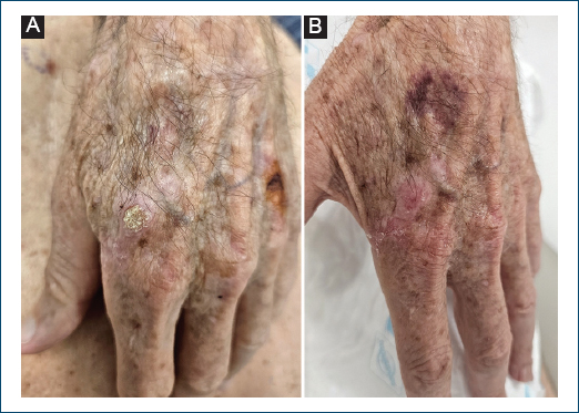
Figure 5. Clinical evolution of a keratoacanthoma on the dorsum of the left hand treated with intralesional methotrexate. A: initial presentation showing a nodular keratotic lesion. B: one month after the second intralesional methotrexate injection, with complete clinical resolution.
All patients were treated with weekly MTX-IL injections and achieved complete clinical resolution. The number of injections required ranged from one to seven, with smaller lesions typically responding after fewer sessions. Progressive necrosis and tumor regression were observed in all cases, with marked improvement typically evident after each treatment session.
In the patient with the bulkiest lesion (Fig. 1A), an initial increase in lesion volume associated with inflammatory signs was observed during the early treatment phase (Fig. 1B); however, this was consistently accompanied by clear clinical evidence of tumor necrosis (Fig. 1C), followed by progressive regression (Fig. 1D and E) and complete resolution (Fig. 1F).
No local or systemic adverse events were recorded. All patients demonstrated good tolerance to the procedure, which was performed without local anesthesia. During follow-up, ranging from 4 months to 12 months, no clinical recurrences were observed.
Discussion
The present series adds to the growing body of evidence supporting MTX-IL as an effective, safe, and minimally invasive treatment for KA. In our cohort, all lesions achieved complete regression without recurrence, aligning with previously reported success rates ranging from 88% to 100%.7–11
The first formal descriptions of MTX-IL for KA date back to Melton et al., who reported complete regression of nine tumors after one to two injections of 0.4–1.5 mL of 12.5–25 mg/mL MTX.12 Subsequent case reports confirmed similar responses in difficult-to-treat anatomical sites such as the nose and lower lip.13,14
A large early dataset reported a 92% complete response across 38 cases treated with MTX-IL, requiring a mean of two injections spaced 2–3 weeks apart. Adverse events were rare, limited to two episodes of pancytopenia in patients with chronic renal failure. This study remains a cornerstone reference, establishing MTX as a credible alternative to surgery for solitary or cosmetically sensitive KAs.7
Further multicenter and prospective experiences, such as a 2014 Korean study (91% clearance across 11 cases treated every 10 days for 2–7 sessions),15 and a 2019 Italian study (100% resolution within 4–8 weeks in 11 patients),16 confirmed the reproducibility of these results. The subsequent 2025 prospective Syrian study treated 37 patients and obtained 94.6% complete regression after four sessions and 98.8% by the eighth, without systemic toxicity.9
Collectively, these data show that most lesions respond after two to five injections of 12.5–25 mg MTX at 1–3-week intervals. Larger or long-standing lesions may require additional sessions, whereas early proliferative KAs typically regress more rapidly.9,15 In our series, the need for a higher number of injections appeared to correlate with lesion volume, whereas no clear association with lesion duration was observed; however, the small sample size precludes any definitive conclusions regarding predictors of response.
A randomized controlled trial directly compared MTX-IL (25 mg/mL) and 5-FU (50 mg/mL), showing complete clearance in 70% and 80% of cases, respectively, with no statistically significant difference.10 The 5-FU group achieved faster resolution (median 2 vs. 3 sessions) but experienced more pronounced local inflammation, whereas MTX showed a more favorable tolerability profile, with less injection-site pain and erythema.
A systematic comparative review of 41 studies corroborated these findings, observing comparable cure rates (92% for MTX and 96% for 5-FU) with a slightly shorter mean healing time for 5-FU (3.7 vs. 4.6 weeks). Both modalities were associated with excellent cosmetic results and negligible systemic toxicity.11
Importantly, both agents demonstrate very low recurrence rates. In a scoping review of 277 patients (336 lesions), 89.4% achieved complete resolution and only 0.8% recurred during a 13-month follow-up.8 Local adverse reactions were mild and self-limited and involved transient injection-site pain in 13% of studies; systemic events were exceedingly rare and largely confined to high-risk individuals with renal impairment.
MTX has also been explored as neoadjuvant therapy, enabling tumor debulking before surgery and improving cosmetic outcomes. Spanish authors demonstrated significant pre-surgical reduction of KA lesion size and scar burden following MTX-IL infusion, with no additional complications.17 This strategy is particularly appealing for large or strategically located tumors where reconstructive morbidity is a concern.
In addition, MTX-IL has also been successfully employed as neoadjuvant therapy in the management of cSCC, achieving significant tumor debulking before definitive surgery. In a retrospective comparative cohort study by Salido-Vallejo et al., 43 patients with histologically confirmed cSCC treated with neoadjuvant MTX-IL showed a measurable reduction in tumor size.18 Among tumors ≥ 2 cm, there was a statistically significant reduction in the number of complex surgical reconstructions required compared with patients undergoing surgery alone. A post hoc analysis revealed that the greatest decrease in tumor size occurred in lesions located on the lower lip. Similarly, in a different prospective study, which included ten patients with cSCC (nine of them located on the lower lip), all lesions decreased in size following MTX-IL, and 30% achieved a complete clinical response.19 Other series have confirmed this pre-operative reduction in tumor dimensions, with ultrasound-based assessments demonstrating decreases not only in tumor diameter but also in thickness after neoadjuvant MTX-IL.20
Overall, the safety profile of MTX-IL is superior to that of systemic retinoids or radiotherapy, and the simplicity of administration facilitates its integration into routine dermatological practice. Nonetheless, caution is warranted in cases of poor renal clearance, where systemic absorption could rarely induce cytopenia.7 Laboratory monitoring – complete blood count and renal and hepatic profiles – is recommended, especially in patients with renal impairment.7,8
Taken together, the literature positions MTX-IL as an excellent therapeutic alternative for KAs in which surgery may result in functional or esthetic compromise, or when systemic comorbidities limit operative management. While surgical excision remains the standard of care, particularly in the context of diagnostic overlap with well-differentiated cSCC, the drug’s mechanism – targeting rapidly dividing keratinocytes through inhibition of dihydrofolate reductase – is particularly suited to the early proliferative phase of KA. From a practical standpoint, MTX offers multiple advantages: simplicity of administration, outpatient feasibility, minimal procedural pain, and negligible cost and risk compared with surgery or radiotherapy. Standard practice involves injecting 12.5–25 mg/mL of MTX (0.3–2 mL) directly into the lesion every 1–3 weeks until blanching occurs, with two to five sessions typically sufficient for complete regression.7,8 Cosmetic outcomes are generally excellent, with post-treatment healing comparable to secondary-intention closure. Reported complications are rare and generally limited to transient local pain, superficial necrosis, or self-limiting ulceration. Systemic absorption is minimal; however, caution is advised in patients with renal or hepatic impairment, in whom reduced drug clearance could theoretically increase the risk of systemic toxicity. Other IL agents, such as 5-FU, may offer similar or even superior efficacy in selected cases, but, in the Portuguese context, the availability and accessibility of MTX – commercially distributed through community pharmacies – make it a more practical and readily implementable therapeutic option for outpatient use.
Limitations of the current evidence include the small size of most studies, heterogeneity of dosing schedules, and limited long-term follow-up. Nevertheless, convergence across independent cohorts – spanning retrospective, prospective, and randomized designs – strongly supports the reproducibility of favorable outcomes. Future multicentre studies should aim to standardize injection protocols, identify predictive factors of response, and define optimal follow-up intervals.
Available data – including the successful outcomes of our own series – confirm that MTX-IL is a safe, effective, and cosmetically favorable therapy for KA, particularly in elderly or surgically ineligible patients. When histopathological confirmation is obtained, MTX should be considered a valid first-line or neoadjuvant option, capable of providing complete clearance with minimal morbidity and excellent patient satisfaction. Its low cost and straightforward outpatient administration make it an attractive alternative to more resource-intensive interventions. Moreover, it may offer the additional advantage of reducing pressure on surgical waiting lists, thereby optimizing the allocation of dermatologic and surgical resources within healthcare systems.
Funding
None.
Conflicts of interest
None.
Ethical considerations
Protection of human subjects and animals. The authors declare that no experiments on humans or animals were performed for this research.
Confidentiality, informed consent, and ethical approval. The authors have followed their institution’s confidentiality protocols, obtained informed consent from all patients, and secured approval from the Ethics Committee. SAGER guidelines have been followed as applicable to the nature of the study.
Declaration on the use of artificial intelligence. The authors declare that no generative artificial intelligence was used in the writing or creation of the content of this manuscript.
