

An otherwise healthy 80-year-old woman presented with a 2-year history of a progressively growing nodule on the scalp, without pain, bleeding, or discharge. The physical examination showed an erythematous-violaceous cupuliform nodule on the left occipital region, measuring 18 × 15 mm (Fig. 1A). No lymphadenopathy was noted. Histopathological examination revealed a circumscribed dermal nodule with pools of mucin separated by thin fibrous septa (Figs. 1B and C), containing islands of neoplastic epithelial cells with eosinophilic cytoplasm, small central nuclei, minimal pleomorphism and no mitotic activity (Fig. 1D), suggestive of primary cutaneous mucinous carcinoma (PCMC). The lesion was excised with clear margins. Extensive diagnostic workup, including breast and pelvic ultrasound, mammography, upper gastrointestinal endoscopy, colonoscopy, maxillofacial, cervical, thorax, abdomen, and pelvis computed tomography ruled out distant primary malignancy. No local recurrence, regional lymph node involvement, or distant metastasis was observed during 2-year follow-up. PCMC is a rare neoplasm arising from eccrine glands and primarily affecting the head and neck area.1 The difficulty of distinguishing this primary neoplasm from metastatic carcinoma of non-cutaneous origins (lung, breast, colon, others) presents a diagnostic challenge.1,2 Microscopically, it is characterized by nests of neoplastic epithelial cells floating in mucinous lakes, with more organized nests, less hyperchromasia, and less mitosis compared to secondary mucinous metastasis. Immunohistochemical markers aid in the diagnosis, but still inconsistently differentiate PCMC from metastatic mucinous adenocarcinomas, so complementary evaluation, such as mammography, gastrointestinal endoscopy, computed tomography, and/or positron emission tomography, should be performed.2–5 This case highlights histological features and diagnostic complexity of PCMC.
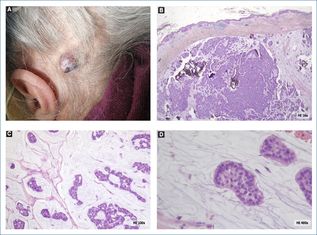
Figure 1. Clinical images. A: elevated, dome-shaped, erythematous-violaceous nodule on the left occipital region; B and C: circumscribed dermal nodule with pools of basophilic mucin divided by thin fibrous septa creating a honeycomb pattern (more evident at right inferior edge); D: within the mucin lakes, “floating” islands of neoplastic epithelial cells with round to cuboidal shape, abundant eosinophilic cytoplasm, small central nuclei, minimal nuclear pleomorphism, and no mitotic figures.
Funding
None.
Conflicts of interest
None.
Ethical considerations
Protection of human subjects and animals. The authors declare that no experiments on humans or animals were performed for this research.
Confidentiality, informed consent, and ethical approval. The authors have followed their institution’s confidentiality protocols, obtained informed consent from all patients, and secured approval from the Ethics Committee. SAGER guidelines have been followed as applicable to the nature of the study.
Declaration on the use of artificial intelligence. The authors declare that no generative artificial intelligence was used in the writing or creation of the content of this manuscript.
