
A 29-year-old woman (Fitzpatrick skin type VI) with no relevant medical history presented to the dermatology emergency department due to a painless, skin-colored papule on her left palm (Fig. 1A). The lesion had been slowly enlarging for about 1 year and was frequently traumatized during her daily activities.
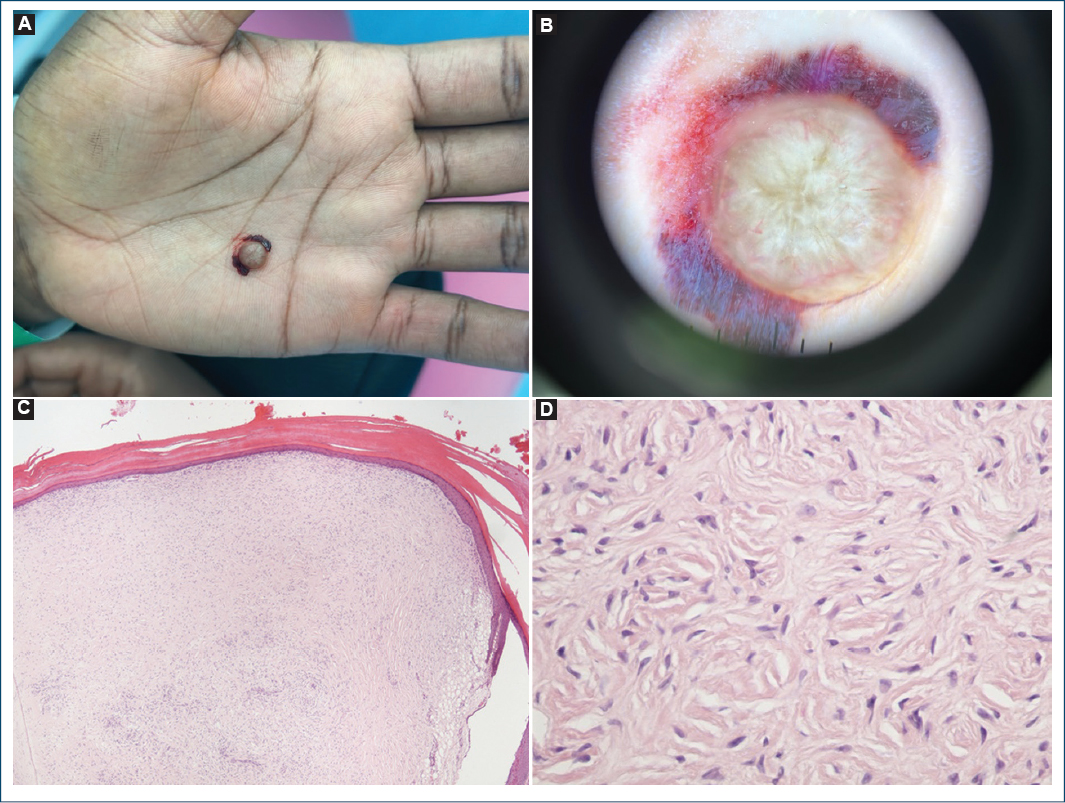
Figure 1. Clinical images. A: solitary skin color papule (palm). B: dermoscopy of the palmar lesion: central homogeneous whitish area with peripheral arborizing vessels and a subtle erythematous halo. C: well-circumscribed but non-encapsulated dermal proliferation composed of thick, paucicellular bundles of hyalinized collage (H&E ×40). D: these randomly oriented clefts, especially at low magnification, confer a storiform appearance (H&E ×100).
On dermoscopic examination (Fig. 1B), a homogeneous whitish area was seen centrally, surrounded by arborizing vessels at the periphery, along with a faint erythematous halo. The lesion was removed by superficial excision (shaving). Histopathology revealed a well-demarcated, hypocellular dermal tumor consisting of thick bundles of hyalinized collagen arranged in a storiform pattern (Fig. 1C), compatible with sclerotic fibroma (SF).
SF, also known as storiform collagenoma, is considered a specific cutaneous marker of Cowden syndrome when multiple lesions are present. However, solitary SFs are not typically associated with this genodermatosis.1 Histopathologically, SF is characterized by a well-circumscribed but non-encapsulated dermal proliferation composed of thick, paucicellular bundles of hyalinized collagen, and traversed by slit-like spaces. These randomly oriented clefts, especially at low magnification, confer a storiform appearance.
Two main theories have been proposed regarding the pathogenesis of SF. The first views SF as a distinct clinicopathological entity, which may explain its association with Cowden syndrome. The second suggests that SF might represent the final sclerotic stage of a preexisting lesion, such as a dermatofibroma, neurofibroma, angiofibroma, folliculitis, erythema elevatum diutinum, lipoma, tendon sheath fibroma, melanocytic nevus, or giant cell collagenoma. Among these possibilities, dermatofibroma is the most commonly associated lesion.2
Dermoscopic features of SF were only recently described. A recent study reported a characteristic pattern composed of a homogeneous white background with peripheral arborizing vessels and, occasionally, an erythematous halo-consistent with our patient’s presentation.1 However, these features are not pathognomonic, and definitive diagnosis relies on histopathological examination and, in selected cases, immunohistochemistry.
In the differential diagnosis, sclerosing perineurioma should be considered, particularly given the unusual palmar location. Other entities include atypical dermatofibroma, late-stage sclerotic dermatofibroma, basal cell carcinoma (which often presents spoke-wheel areas, maple leaf-like structures, ulceration, or blue-gray ovoid nests), and hypomelanotic or amelanotic blue nevus and melanoma.3
Immunohistochemistry was performed in our case. The lesion was CD34-positive, Factor XIIIa-positive, and epithelial membrane antigen-weakly positive. This profile also supported the diagnosis of SF.
Most cases of SF are solitary and not associated with systemic disease. However, when multiple lesions are present, Cowden syndrome should be ruled out2. Surgical excision is generally performed for cosmetic reasons or due to recurrent trauma, with an excellent prognosis and low recurrence rates.2,3
Acknowledgments
We would like to thank Dr. P. de Vasconcelos for providing the histological photographs.
Funding
None.
Conflicts of interest
None.
Ethical considerations
Protection of humans and animals. The authors declare that no experiments involving humans or animals were conducted for this research.
Confidentiality, informed consent, and ethical approval. The authors have followed their institution’s confidentiality protocols, obtained informed consent from patients, and received approval from the Ethics Committee. The Sex and Gender Equity in Research guidelines were followed according to the nature of the study.
Declaration on the use of artificial intelligence. The authors declare that no generative artificial intelligence was used in the writing of this manuscript.
