Apocrine hidrocystoma of the breast: a case report of unusual clinical and dermoscopic findings

Apocrine hidrocystoma of the breast: a case report of unusual clinical and dermoscopic findings

Ma. Cristina Fialho 1 , Emanuel Marques 1, 2 , Monika Arenbergerová 3, 4 , Petr Arenberger 3, 4 , Zoltán Paluch 5, 6 , Marek Pásek 3, 4

1 Department of Dermatology and Venereology, Hospital de Santo António dos Capuchos, Unidade Local de Saúde de São José, Lisbon, Portugal; 2 Department of Dermatology and Venereology, NOVA Medical School, NOVA University of Lisbon, Lisbon, Portugal; 3 Department of Dermatology and Venereology, Královské Vinohrady University Hospital, Prague, Czech Republic; 4 Department of Dermatology and Venereology, Third Faculty of Medicine, Charles University, Prague, Czech Republic; 5 Department of Clinical Pharmacology and Pharmacy, General University Hospital, Prague, Czech Republic; 6 Department of Clinical Pharmacology and Pharmacy, General University Hospital, Institute for the Care of Mother and Child, Prague, Czech Republic

Ma. Cristina Fialho, Emanuel Marques, Monika Arenbergerová, Petr Arenberger, Zoltán Paluch, Marek Pásek

La información completa de afiliaciones y autor de correspondencia está disponible en la versión original en PDF.

*Correspondence: Ma. Cristina Fialho. Email: cris.fialho.3@gmail.com

Ma. Cristina Fialho1*thumb, Emanuel Marques1,2thumb, Monika Arenbergerová3,4thumb, Petr Arenberger3,4thumb, Zoltán Paluch5,6thumb, Marek Pásek3,4thumb

1Department of Dermatology and Venereology, Hospital de Santo António dos Capuchos, Unidade Local de Saúde de São José, Lisbon, Portugal; 2Department of Dermatology and Venereology, NOVA Medical School, NOVA University of Lisbon, Lisbon, Portugal; 3Department of Dermatology and Venereology, Královské Vinohrady University Hospital, Prague, Czech Republic; 4Department of Dermatology and Venereology, Third Faculty of Medicine, Charles University, Prague, Czech Republic; 5Department of Clinical Pharmacology and Pharmacy, General University Hospital, Prague, Czech Republic; 6Department of Clinical Pharmacology and Pharmacy, General University Hospital, Institute for the Care of Mother and Child, Prague, Czech Republic

*Correspondence: Ma. Cristina Fialho E-mail: cris.fialho.3@gmail.com

Received: 28-03-2025
Accepted: 09-04-2025
DOI: 10.24875/PJDV.25000022
Available online: 25-05-2025
Port J Dermatol and Venereol. 2026;84(2):124-125

Contents

A 33-year-old woman presented with a 6-month history of a gradually increasing papule on her right breast. A 17 mm red-violet, asymptomatic, relatively indurated dome-shaped nodule was identified (Fig. 1A). Dermoscopic examination (Fig. 1B) revealed a central homogeneous bluish-gray area with a few short linear-­irregular vessels, an erythematous area with shiny whitish structures and a pigmented pseudo-network. Ultrasound examination revealed a cystic hypoechoic mass. Excision of the lesion was proposed, and the patient was at her 36th week (+4 days) of pregnancy on the day of the procedure. Histologic examination revealed an apocrine hidrocystoma.
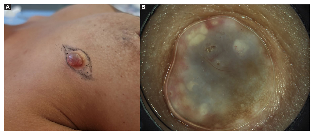
Figure 1. Clinical images. A: clinical picture of the dome-shaped nodule on the right breast; B: dermoscopy of the apocrine hidrocystoma in vivo before excision.

Apocrine hidrocystomas are benign tumors of the apocrine sweat glands most frequently found on the head and neck.1 Few have been reported in other areas.14 Most grow to sizes varying from 1 to 10 mm; there are only few reports of larger lesions.2 Our dermoscopic findings do not correspond to the most frequent patterns reported by Zaballos et al.1 gray and pink homogenous areas and linear-irregular vessels were only found in 4.8% and 9.1% of reported cases; moreover, pigmented pseudo-networks have not been described.1 In the differential diagnosis, atypical dermatofibroma, pigmented basal cell carcinoma, amelanotic melanoma or other adnexal tumors should be considered, particularly when atypical dermoscopic findings are present.3 We hypothesize that a pigmented pseudo-network may have been caused by irritation derived from a chronic inflammatory process. The atypical findings of our hidrocystoma enhance the need for physicians to be aware of uncommon locations and dermoscopic findings of these lesions.

Funding

None.

Conflicts of interest

None.

Ethical considerations

Protection of human subjects and animals. The authors declare that no experiments on humans or animals were performed for this research.

Confidentiality, informed consent, and ethical approval. The authors have followed their institution’s confidentiality protocols, obtained informed consent from all patients, and secured approval from the Ethics Committee. SAGER guidelines have been followed as applicable to the nature of the study.

Declaration on the use of artificial intelligence. The authors declare that no generative artificial intelligence was used in the writing or creation of the content of this manuscript.

References

1. Zaballos P, Bañuls J, Medina C, Salsench E, Serrano P, Guionnet N. Dermoscopy of apocrine hidrocystomas:a morphological study. J Eur Acad Dermatol Venereol. 2014;28:378-81.

2. May C, Chang O, Compton N. A giant apocrine hidrocystoma of the trunk. Dermatol Online J. 2017;23:13030/qt8bm3r2h4.

3. Cieza-Díaz DE, Avilés-Izquierdo JA, Ciudad-Blanco C, Parra-Blanco V. Dermatoscopy of apocrine tumors:report of 4 cases. Actas Dermosifiliogr. 2013;104:826-8.

4. Samplaski MK, Somani N, Palmer JS. Apocrine hidrocystoma on glans penis of a child. Urology. 2009;73:800-1.