

Dear Editor,
A 60-year-old woman with a prior right radical nephrectomy for clear cell renal carcinoma was admitted 18 months later with a 5-day history of expressive aphasia and right-sided motor weakness. During hospitalization, dermatology evaluated a 1.5 cm erythematous-violaceous nodule on the right flank, soft in consistency, evolving over several months (Fig. 1). Excisional biopsy revealed a dermal-based nodule with central necrosis and epithelioid cells with moderate atypia (Fig. 2). Immunohistochemistry was positive for CK AE1/AE3 and vimentin, consistent with cutaneous metastasis of renal origin. Concurrent staging with computed tomography and positron emission tomography (PET) identified a left frontal infra-axial lesion suggestive of brain metastasis, pulmonary nodules likely secondary, and probable peritoneal involvement. PET imaging demonstrated radiological-clinical correlation with the cutaneous lesion (Fig. 3).
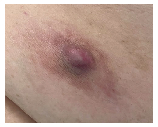
Figure 1. Erythematous-violaceous nodule on the right flank.
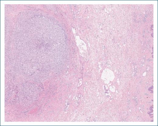
Figure 2. Histology showing a dermal-based nodule with extensive central necrosis and epithelioid cells exhibiting moderate atypia (H&E ×2).
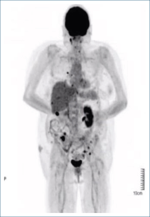
Figure 3. PET scan showing a hypermetabolic lesion in the right flank (circled), corresponding to a confirmed metastasis. PET: positron emission tomography.
Surgical excision confirmed brain metastasis from renal cell carcinoma. She received radiotherapy to the surgical bed and palliative sunitinib but died 5 months after starting treatment.
Cutaneous metastases from renal cell carcinoma are a manifestation of advanced disease, typically associated with poor prognosis, and may represent the first sign of metastatic spread.1 Clinically, they show varied presentations and may mimic various benign and malignant lesions, including hemangioma, pyogenic granuloma, angiosarcoma, and Kaposi sarcoma.1,2 Surgical excision is the preferred treatment for solitary lesions.3 However, up to 90% of cases occur with visceral metastases, explaining the poor prognosis and median survival under 6 months.3
As observed in this case, the cutaneous nodule preceded cerebral metastasis, and earlier detection might have influenced both prognosis and therapeutic approach.
Funding
None.
Conflicts of interest
None.
Ethical considerations
Protection of human subjects and animals. The authors declare that no experiments on humans or animals were performed for this research.
Confidentiality, informed consent, and ethical approval. The authors have followed their institution’s confidentiality protocols, obtained informed consent from all patients, and secured approval from the Ethics Committee. SAGER guidelines have been followed as applicable to the nature of the study.
Declaration on the use of artificial intelligence. The authors declare that no generative artificial intelligence was used in the writing or creation of the content of this manuscript.
