

Introduction
Seborrheic keratosis (SK), solar lentigo (SL), pigmented actinic keratosis (pAK), and lichenoid keratosis (LK) are common benign skin lesions. These lesions often appear near each other or may develop from one another. Pigmented facial lesions frequently present to physicians and can be challenging to distinguish because the differential diagnosis includes a wide range of benign and malignant conditions with similar clinical appearances, particularly in dark skin phototypes. In many cases, diagnostic uncertainty is not resolved by clinical inspection or by dermatoscopy. This usually leads to confocal microscopy, biopsy, or excision to rule out melanoma in situ.1
On the other hand, flat pigmented lesions on the face are a common reason for consultation, with patients requesting rapid, non-surgical treatment by cryotherapy, laser, or other local treatment modalities, sometimes without a definitive histopathological diagnosis. This procedure carries the risk of inappropriate treatment of melanoma and a delayed diagnosis, which once again highlights the importance of dermoscopy.2
Dermoscopy is a non-invasive clinical method that is crucial for diagnosing and managing pigmented skin lesions in comparison with examination with the unaided eye.
Our study aimed to describe the epidemiological profile and identify the most frequent and specific dermoscopic signs of solar lentigines, flat SK, LK, pAK, and finally malignant lentigo, especially in dark skin phototypes.
Materials and methods
Study design and patients
We conducted a retrospective and prospective observational study involving 406 lesions from 274 patients diagnosed in our department.
- – The inclusion criteria comprised all patients presenting with acquired flat pigmented lesions of the face
- – The exclusion criteria included infiltrated lesions, ephelides, melanocytic nevi, basal cell carcinomas, and flat pigmented lesions located outside the facial area.
Patients were recruited from outpatient consultations, inpatient hospital activity, and referrals to the dermatology department.
A Dermlite DL4 and DL5 dermoscopes were used with ×10 magnification, both with and without immersion fluid, either hand-cleansing gel containing 70% ethanol or ultrasound gel with and without polarized light, and with the pigment boost option combined with an iPhone 11 smartphone. All patients underwent a clinical and dermoscopic examination, after which photographs were taken for each lesion and subsequently evaluated by two different dermatologists who were blinded to the histopathological diagnosis. Data collection was carried out using a structured data processing form and recorded in an Excel file. The following parameters were evaluated:
Clinical data: age, sex, Fitzpatrick skin type, and localization (cheeks, forehead, periorbital region, nose, ears, and mandibular area).
The diagnoses of SL, SK, pAK, and LK were established based on clinical and dermoscopic criteria described in the literature (Table 1).
Table 1. Definitions of the dermoscopic patterns examined in our study
| Lesion | Dermoscopic pattern | Definition |
|---|---|---|
| SL | Structureless areas | Many lesions lack specific dermoscopic structures or a pigment network and present only light brown homogeneous (structureless) areas.3 |
| Moth-eaten border | Irregularly curved and sharply demarcated edge.3 | |
| Fingerprint-like areas | Fine, parallel lines resembling dermatoglyphics are caused by elongated epidermal ridges.3 | |
| Jelly sign | Superficial pigmentation extending in a liquid-like manner around follicular openings.3 | |
| Pseudonetwork | Facial pigmentation is interrupted by follicular and adnexal openings.3 | |
| SK | Fissures | Brown linear or curvilinear structures that may coalesce into a cerebriform pattern; histologically correspond to deep epidermal invaginations filled with keratin.4 |
| Comedo-like openings | Round to oval keratin-filled clefts, usually dark brown, gray, or black; histologically correspond to concave epidermal invaginations filled with keratin.4 | |
| Fat finger | Thick, gently curved lines, sometimes showing branching, with coloration ranging from hypopigmented to brown, black, or blue. The term “fat finger” is typically applied to flat or slightly elevated early seborrheic keratosis.4 | |
| pAK | Rhomboidal pattern | A grayish-brown pigmented pseudonetwork forming rhomboidal structures around follicular openings; histologically reflects hyperpigmentation of the Malpighian layer interrupted by follicles.5 |
| Scales and crusts | Superficial scales and crusts, which are not obligatorily centered within the follicular unit. | |
| Rosettes | Four bright white dots arranged in a clover-like configuration, predominantly within follicular openings; correspond histologically to orthokeratosis and parakeratosis (“keratin pearls”), reflecting intraepidermal keratin whorls or horn pearls. | |
| LK | Annular granular pattern | Grayish-brown dots and globules arranged annularly around hair follicles; histologically correspond to clusters of melanin and melanophages within the papillary dermis.6 |
|
SL: solar lentigo; SK: seborrheic keratosis; LK: lichenoid keratosis; pAK: pigmented actinic keratosis; LM: lentigo maligna. |
||
Diagnosis
The diagnosis was based on clinical and dermoscopic evaluation. Histopathological confirmation was obtained whenever required for diagnostic clarification. Informed consent was obtained from all subjects involved in the study. The study was conducted in accordance with the principles outlined in the Declaration of Helsinki.
Statistical analysis
Statistical analysis was conducted using the Statistical Package for the Social Sciences 20.0 software. Descriptive and analytical comparisons of dermoscopic findings were conducted. Dermoscopic features were compared across lesion types using Fisher’s exact test to determine statistical significance (p < 0.05).
Results
Patient analysis
The study included 406 lesions from 274 patients. The mean age was 62.9 ± 10.7 years (range: 30–82 years) with a median of 65 years. The study population comprised 61.8% women and 38.2% men, with a sex ratio of 0.62. Phototype IV was predominant (60.2%), followed by phototype III (33.3%). Regarding anatomical location, the cheek was the most common site (59.9%), followed by the forehead (23.6%), the periorbital region and nose (7%), and the ears and mandibular area (1.3%).
Among the 406 analyzed cases, in SL (n = 170), the most frequent dermoscopic features were dermoscopic lightening (78.3%, p = 0.012), sharp borders (70.6%, p = 0.018), and homogeneous brown pigmentation (67.3%, p = 0.021). Moth-eaten borders were also significantly associated with this lesion type (57%, p = 0.034), as was symmetrical perifollicular pigmentation (37.8%, p = 0.029) (Fig. 1A).
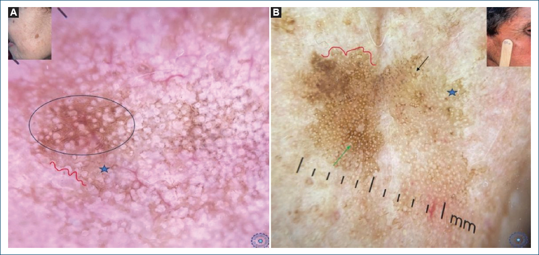
Figure 1. Clinical images. A and B: dermoscopic features of solar lentigo showing fingerprint-like areas (blue circle), jelly sign (blue star), and moth-eaten border (red line), and symmetric pseudonetwork (green arrow).
In contrast, fingerprint-like structures (41.4%, p = 0.087), brown pseudonetwork (30.7%, p = 0.094), and asymmetrical perifollicular pigmentation (7.2%, p = 0.214) did not show a statistically significant association (Fig. 1B). SK (n = 83) was significantly associated with sharp borders (75.5%, p = 0.009), fingerprint pattern (57%, p = 0.015), fissures and ridges (45%, p = 0.022), pseudocomedones (26.5%, p = 0.031), and cerebriform pattern (24%, p = 0.040) (Fig. 2).
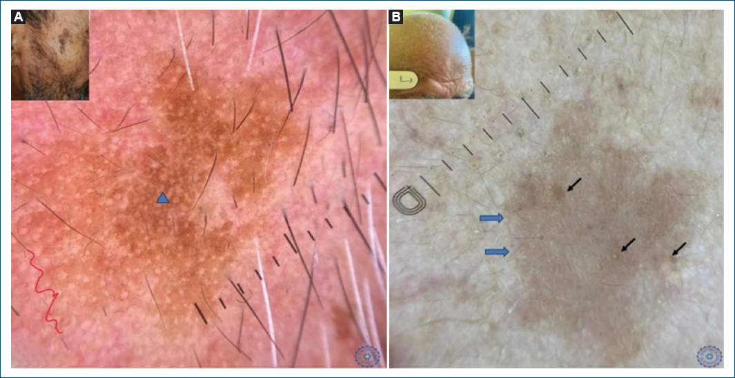
Figure 2. Clinical images. A and B: dermoscopic features of seborrheic keratosis showing fissures and ridges forming a cerebriform pattern (blue triangle), sharp border (blue arrow), moth-eaten border (red line), and pseudocomedones (black arrow).
In LK (n = 60), a diffuse granular pattern was the predominant and significantly associated feature (80%, p = 0.006). The localized granular pattern (20%, p = 0.118) did not reach statistical significance (Fig. 3).
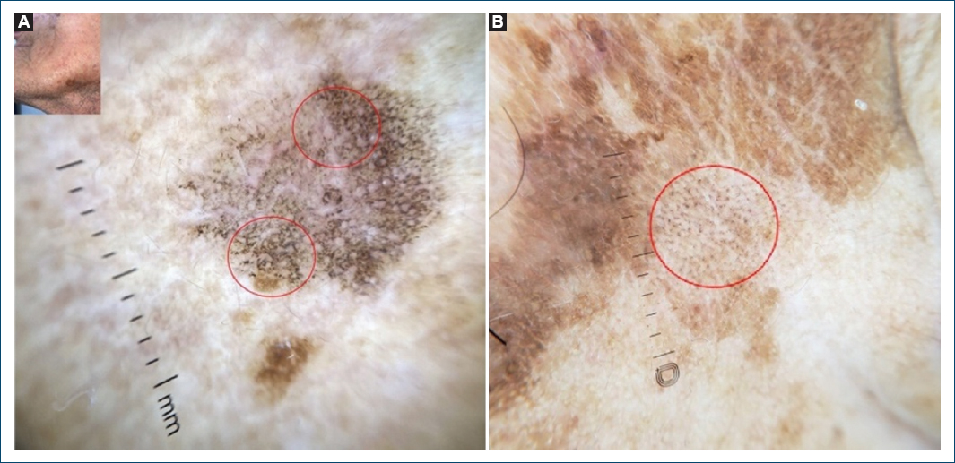
Figure 3. Diffuse granular pattern (A and B: red circles).
pAK (n = 82) was mainly characterized by a pigmented pseudonetwork (70%, p = 0.014). Other significantly associated features included the rhomboidal pattern (26%, p = 0.028), gray halos (15%, p = 0.033), and annular granular pattern (17%, p = 0.025). Yellow follicles (18%, p = 0.176) were not significantly associated (Fig. 4).
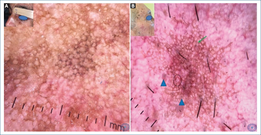
Figure 4. Dermoscopic features of pigmented actinic keratosis showing rhomboidal pattern (A), gray halo (blue triangle), rosettes (dark circle), and symmetric pseudonetwork (green arrow) (B).
Finally, in lentigo maligna (LM) (n = 11), asymmetrical pigmented follicular openings were the most strongly associated feature (75%, p = 0.011), followed by target-like appearance (62%, p = 0.019) and rhomboidal structures (43%, p = 0.037) (Fig. 5). Gray coloration (39%, p = 0.082) was observed but did not show statistical significance (Table 2).
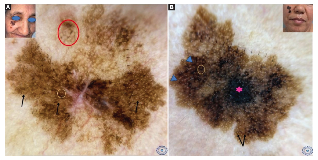
Figure 5. Clinical images. A and B: dermoscopic features of lentigo maligna showing rhomboid structures (blue triangle), asymmetric pigmented follicular openings and atypical pseudonetwork (black arrow), dark blotches (pink star), target-like appearance (yellow circles), and granular and annular pattern (red circle).
Table 2. Analysis results of dermoscopic feature frequency according to lesion type and statistical significance
| Lesion type | Dermoscopic feature | Frequency (%) | p |
|---|---|---|---|
| SL (n = 170) | Dermoscopic lightening | 78.3 | 0.012 |
| Sharp border | 70.6 | 0.018 | |
| Homogeneous brown pigmentation | 67.3 | 0.021 | |
| Moth-eaten border | 57 | 0.034 | |
| Symmetrical perifollicular pigmentation | 37.8 | 0.029 | |
| Fingerprint-like structures | 41.4 | 0.087 | |
| Brown pseudonetwork | 30.7 | 0.094 | |
| Asymmetrical perifollicular pigmentation | 7.2 | 0.214 | |
| SK (n = 83) | Sharp border | 75.5 | 0.009 |
| Fingerprint pattern | 57 | 0.015 | |
| Dermoscopic lightening | 59.2 | 0.001 | |
| Fissures and ridges (fat fingers) | 45 | 0.022 | |
| Moth-eaten border | 44.9 | 0.059 | |
| Brown pseudonetwork | 30.7 | 0.986 | |
| Pseudocomedones | 26.5 | 0.031 | |
| Cerebriform pattern | 24 | 0.040 | |
| LK (n = 60) | Diffuse granular pattern | 80 | 0.006 |
| Dermoscopic lightening | 36 | 0.005 | |
| Localized granular pattern | 20 | 0.118 | |
| Brown pseudonetwork | 17.2 | 0.005 | |
| pAK (n = 82) | Pigmented pseudonetwork | 70 | 0.014 |
| Rhomboidal pattern | 26 | 0.028 | |
| Gray halos | 15 | 0.033 | |
| Annular granular pattern | 17 | 0.025 | |
| Yellow follicles | 18 | 0.176 | |
| LM (n = 11) | Asymmetrical pigmented follicular openings | 75 | 0.011 |
| Target-like appearance | 62 | 0.019 | |
| Rhomboidal structures | 43 | 0.037 | |
| Gray coloration | 39 | 0.082 | |
|
SL: solar lentigo; SK: seborrheic keratosis; LK: lichenoid keratosis; pAK: pigmented actinic keratosis; LM: lentigo maligna. |
|||
Discussion
SK, SL, pAK, and LK, also termed lichen planus-like keratosis, are benign non-melanocytic lesions that frequently coexist in the same individual and may be difficult to differentiate, despite each having well-defined clinical and dermoscopic characteristics.1 Histopathologically, these entities are distinct; however, because of their benign nature, biopsies are generally performed only in cases where melanoma is suspected.7
In clinical practice, dermoscopy is widely recognized as a valuable non-invasive tool that significantly improves the diagnostic accuracy of pigmented skin lesions compared to visual inspection alone.8
Solar lentigines are among the most common benign pigmented skin lesions, typically presenting as well-defined, uniformly pigmented macules on sun-exposed areas. They may occasionally mimic LM, which often results in an excessive number of biopsies performed on benign lesions, a practice that dermoscopy can help reduce. Indeed, this phenomenon of “over-biopsy,” driven by the fear of missing an early primary melanoma, represents a scenario in which dermoscopy is particularly valuable.
The main objective of this study was to evaluate and compare the dermoscopic characteristics of common facial pigmented lesions in individuals with dark phototypes, and to determine their diagnostic relevance for SL, SK, LK, pAK, and LM. Existing literature on this topic focuses primarily on fair skin phototypes, highlighting the need for dermoscopic data specific to dark skin phototypes.
In our series, the frequency of SL-specific dermoscopic criteria was comparable to that reported by Scott and Oakley, particularly homogeneous brown pigmentation, sharply demarcated borders, and the so-called “enlightenment” (lightening of pigmentation). However, cases of asymmetric follicular pigmentation were observed in our patients (7.2%), although this was not statistically significant (p > 0.05).9
Flat seborrheic keratoses typically present as oval macules ranging from light to dark brown with well-defined borders. As they progress, they evolve into plaques with a waxy, “stuck-on” appearance. Dermoscopy is particularly useful in atypical or irritated presentations and in early superficial lesions, as in our series. In addition to the moth-eaten borders and fingerprint-like structures described in SL, the maturation of these lesions revealed fissures and comedo-like openings.
In contrast to Scott and Oakley’s series, SK in our patients showed darker brown dots and granules, a difference likely attributable to the darker phototypes of our population. Moreover, dermoscopic features typically associated with SL (lightening, jelly sign, moth-eaten borders, and brown pseudonetwork) were also observed in seborrheic keratoses, further reinforcing their overlap.9
LK may appear as macular or slightly infiltrated lesions, with brown, gray, or erythematous coloration. It results from an immunologic or inflammatory reaction to a pre-existing lesion, such as SK or SL. The hallmark dermoscopic feature is a granular pattern, consisting of brown, gray, or bluish granules with a localized or diffuse distribution, corresponding histologically to pigment incontinence and melanophages in the papillary dermis.9 Unlike our findings, Rebecca’s series showed a predominance of the localized annular-granular pattern, probably due to a higher prevalence of erythematous LK.9 Nevertheless, residual dermoscopic features of SK and SL observed in both studies support the concept of an evolutionary link among these lesions.
In our series, pAK was primarily characterized by the presence of a pigmented pseudonetwork, frequently accompanied by annular-granular structures, a gelatinous appearance, gray halos, and rhomboidal patterns.
These observations are partially consistent with published data. Kelati et al. described the same patterns associated with scaling and superficial pigmentation, suggesting a more significant follicular and keratinous component than that observed in our cohort.5
Similarly, Akay et al. identified slate-gray dots, brown-gray pseudonetworks, and rhomboidal structures as frequent dermoscopic signs of pigmented facial lesions, highlighting the relative specificity of the pseudonetwork for pAK.10 Our results confirm this observation, with the pseudonetwork constituting a central dermoscopic feature in our series.
However, contrary to the data in the literature, Tschandl et al. and Lallas et al. reported that gray halos and circles were more strongly associated with LM than with pAK.11,12 In our study, although gray halos were observed in pAK, they were not the predominant feature, which could explain their greater diagnostic relevance for LM than for pAK.
Early clinical diagnosis of LM remains challenging, as it can closely mimic SL or inflamed SK, particularly when multiple lesions coexist. Dermoscopic differentiation is useful: pseudofollicular openings, milia-like cysts, opaque yellowish homogeneous areas, cerebriform structures, and fingerprint-like patterns are more commonly observed in benign pigmented facial lesions. In contrast, asymmetric pigmented follicular openings, rhomboidal structures, dark streaks, dark globules, and dots are more characteristic of malignant pigmented facial lesions.13
In our series, LM was predominantly characterized by asymmetric pigmented follicular openings (75%, p = 0.011) and target-like structures (63%, p = 0.019). In contrast, studies conducted in turkish cohorts reported a higher diagnostic relevance of rhomboidal structures, and black blotches (75%).13 The prevalence of grey areas appeared comparable between the two populations (39%).
It is important to emphasize that asymmetric pigmented follicular openings are not exclusively specific to melanoma. Although considered a key diagnostic clue for LM, this feature may also be observed in benign pigmented lesions. In our cohort, asymmetric perifollicular pigmentation was identified in a subset of solar lentigines (7.2%, p = 0.214). This overlap underlines the need for cautious interpretation of follicular criteria, particularly in populations with darker phototypes where pigment distribution may be more heterogeneous.
Limitations
The limitations of our study include its single-center design, the relatively small number of LM cases, and the absence of systematic histopathological confirmation for all lesions.
Conclusion
The dermoscopic characteristics of pigmented lesions on sun-exposed skin largely overlap and can vary according to phototype, hence the importance of dermoscopic analysis according to phototype.
Funding
None.
Conflicts of interest
None.
Ethical considerations
Protection of human subjects and animals. The authors declare that no experiments on humans or animals were performed for this research.
Confidentiality, informed consent, and ethical approval. The authors have followed their institution’s confidentiality protocols, obtained informed consent from all patients, and secured approval from the Ethics Committee. SAGER guidelines have been followed as applicable to the nature of the study.
Declaration on the use of artificial intelligence. The authors declare that no generative artificial intelligence was used in the writing or creation of the content of this manuscript.
