

Introduction
Erythema multiforme (EM), an immune-mediated hypersensitivity reaction, is an affection of the skin and mucous membranes.1 It results in macular, papular, urticarial, or bullous lesions, typically presenting as classic “target lesions” mainly in the extremities.2 Affected mucous membranes (oral, ocular, and/or genital) are more commonly observed in the EM major than the minor form.1 Infections are the most frequent causes, with herpes simplex virus (HSV) playing the major role.3 Varicella-zoster virus (VZV) is a rare trigger,4 and its association with primary varicella is uncommon, with only a few documented cases.5–7 We report a case of a 44-year-old woman who developed EM major 1 week after the onset of typical skin lesions of varicella.
Case report
A 44-year-old female patient was admitted to our hospital with a widespread rash involving the whole body, which was composed of lesions in different stages, such as erythematous macules, papules, and translucent vesicles, some of them umbilicated, on the forearms and trunk that had been lasting for 3 days (Fig. 1). The skin rash was preceded by pain and fever. During history-taking, the patient denied any prior episode of varicella in childhood. The diagnosis of primary varicella was made clinically based on prodromal symptoms, a characteristic polymorphic rash with lesions at different stages starting on the trunk and scalp, and centrifugal spread. Laboratory workup (complete blood count, liver and renal function tests) and chest X-ray were normal. Five days later, she developed new lesions and was readmitted for further assessment. There was no history of previous medication. On dermatological examination, there were multiple erythema target lesions with erosive centers, some of which had purpuric areas and desquamation, diffusely distributed over the body, associated with a few clear vesicles (Fig. 2). The buccal mucosa showed mild erosions and dry cheilitis, but no genital lesions were seen, nor was ocular involvement present. The Nikolsky sign was negative. A diagnosis of EM associated with varicella was made based on clinical symptoms and confirmed by skin biopsy, which demonstrated epidermal hyperkeratosis and focal subepidermal detachment with rare apoptotic bodies, spongy edema, and infiltration of lymphocytes (Fig. 3). The search for immunosuppression was negative, and the patient’s condition improved with supportive care, topical corticosteroids, and a 7-day course of acyclovir. At the 2-month follow-up after discharge, the patient reported complete resolution of the rash with no recurrence; examination found only residual hyperpigmented macules.
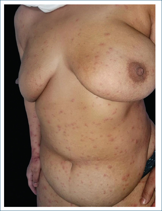
Figure 1. Vesiculo-maculopapular eruption on the trunk and right upper extremity.
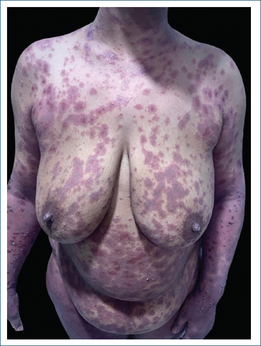
Figure 2. Target or iris lesions on the trunk and extremities with hemorrhagic crusts in places.
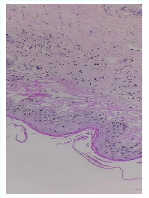
Figure 3. The biopsy specimen on the right thigh shows a scattered lymphocytic infiltration and vacuolar degeneration and necrotic keratinocyte (H&E staining, 200×).
Discussion
EM was first described by Ferdinand von Hebra in 1866.8 It can affect people of any age, though it most often appears between 20 and 40 years, with a slight male predominance2 and an estimated annual incidence of < 1%. in the general population.3 Clinically, EM is recognized by papules that evolve into the classic target or iris lesions. These have at least three concentric zones with well-defined borders that can appear as macular, papular, vesicular, or even bullous. They usually show up within 48–72 h.2 The rash starts symmetrically on the extremities, mainly on extension surfaces,3 then can spread more widely across the body and sometimes take on a purpuric appearance.9 The condition generally resolves within 1 week to 21 days.2 The histopathological features of EM include a prominent lymphocytic infiltrate, predominantly composed of T-lymphocytes, along the dermoepidermal junction and around superficial dermal vessels, accompanied by dermal edema, epidermal keratinocyte necrosis, and subepidermal bullae formation.10 A variety of etiologic factors have been reported to cause EM, including medications, autoimmune disorders, and vaccinations.2 However, infectious agents are a major cause, with HSV-1 in 66.7% of cases, HSV-2 in 27.8%.3 Although VZV infection has rarely been associated with EM, our literature review identified only a few case reports linking the two,4–7,9–13 most of which involved herpes zoster rather than primary varicella. Notable cases of EM secondary to primary varicella have been described in the literature, though they remain uncommon, particularly in adults. For instance, Kishore et al.6 reported a 14-year-old girl who developed EM 2 weeks after a varicella rash, presenting with vesicles on an erythematous base and target lesions involving the trunk, extremities, face, and scalp; the diagnosis was confirmed by VZV polymerase chain reaction, and she was treated with intravenous acyclovir. In a similar vein, Liyanage et al.7 described a 35-year-old man with a 3-day history of generalized erythematous vesicular rash that progressed to targetoid lesions and oral vesicles; biopsy confirmed EM, with a Tzanck smear supporting varicella. Our case closely resembles these reports, demonstrating EM in an immunocompetent adult following primary varicella, biopsy-proven, and responding well to oral acyclovir. In contrast, the majority of documented cases involve EM triggered by herpes zoster reactivation rather than primary varicella, and this appears more common in adults. Weisman et al.11 described four patients aged 20–95 years who developed bullous or target lesions days to weeks after thoracic herpes zoster; diagnoses were made clinically or by biopsy, with VZV serology or antigen detection, and disease duration ranged from 7 days to 3 weeks with a fatal outcome in one elderly patient. Other reports follow a similar pattern: Wollina and Gemmeke10 documented a 76-year-old man with widespread EM after thoracic zoster, biopsy-confirmed and resolving over 2 weeks; Zhu et al.4 presented a 50-year-old man whose EM appeared 9 days after herpes zoster on the left thorax, with biopsy showing lymphocytic infiltrate. He was treated successfully with oral acyclovir and topical mometasone. Additional cases, such as those by Park et al.12 and Aljehani et al.13, show targetoid or annular lesions developing 3–10 days after zoster onset, often biopsy-confirmed and responding favorably to supportive care with antivirals. Prais et al.5 also highlighted pediatric cases associated with varicella and Ramsay-Hunt syndrome, with durations varying up to 8 months. Overall, EM triggered by primary varicella seems distinctly less frequent in adults compared with zoster-associated cases. When it occurs, the onset is typically 3–14 days after the viral rash, and outcomes are generally favorable with antiviral therapy and supportive measures. It’s very important to distinguish the vesicular lesions that show up as part of the polymorphous rash in EM from the classic vesicles seen in VZV infection. Given how common chickenpox and herpes zoster actually are, it’s striking that there are still only a rare reported cases linking VZV to EM. One possible explanation is that when EM already has these central vesicles as one element of its varied eruption, it can easily hide or confuse the typical appearance of primary varicella. Acute EM is managed supportively in most cases, with treatment aimed at relieving symptoms and providing comfort.2 Antivirals are added when an active viral cause is confirmed.3 In our patient, the active lesions cleared within 7 days, and at the 2-month follow-up, she had only residual hyperpigmented macules remaining. This case brings another example to the very small body of evidence linking primary varicella to EM and reminds us to keep VZV in mind when dealing with post-infectious presentations.
Conclusion
The rarity of varicella-associated erythema multiforme in adults, as evidenced by limited historical cases, calls for a reevaluation of its diagnostic thresholds in clinical practice. This underscores the need for heightened awareness among healthcare providers to ensure timely recognition and management, paving the way for targeted research to address this understudied condition.
Funding
None.
Conflicts of interest
None.
Ethical considerations
Protection of human subjects and animals. The authors declare that no experiments on humans or animals were performed for this research.
Confidentiality, informed consent, and ethical approval. The authors have followed their institution’s confidentiality protocols, obtained informed consent from all patients, and secured approval from the Ethics Committee. SAGER guidelines have been followed as applicable to the nature of the study.
Declaration on the use of artificial intelligence. The authors declare that no generative artificial intelligence was used in the writing or creation of the content of this manuscript.
