

An 18-month-old girl was evaluated for pruritic facial lesions evolving over 2 months. She was healthy, with no personal or family dermatological history. She frequently visited her grandmother’s farm, where she had contact with chickens and rabbits. Examination revealed a symmetrical, erythematous butterfly-shaped plaque occupying the central facial region, extending to the glabella and supraorbital areas, covered by yellowish scales (Fig. 1). The parents reported prior application of methylprednisolone aceponate cream, which worsened the dermatosis.
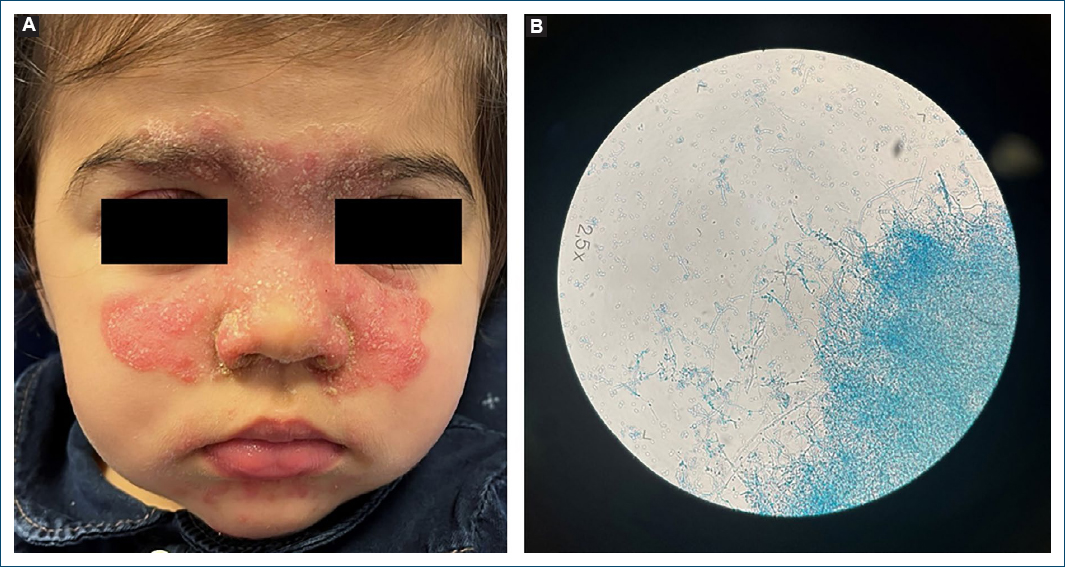
Figure 1. Clinical images. A: clinical findings showing erythematous butterfly-shaped plaque on the central facial region, covered by yellowish scales; B: microscopic examination of culture specimens identified clusters of microconidia and spiral hyphae, consistent with Trichophyton mentagrophytes (var. granulosum).
What is the diagnosis?
Tinea incognita
A mycological examination showed septate filaments on direct microscopy. Microscopic examination of culture specimens identified clusters of microconidia and spiral hyphae, consistent with Trichophyton mentagrophytes (var. granulosum). The diagnosis of tinea incognita (TI) was established.
TI is a dermatophytosis altered by topical corticosteroids or calcineurin inhibitors, masking its clinical features.1 It commonly affects children aged 10-14 years and is rare in those under 3 years.2,3 The face is frequently involved, as tinea faciei can be diagnostically challenging.2,3 Hyphal invasion of hair follicles often necessitates oral antifungal treatment.
T. mentagrophytes var. granulosum is a zoophilic dermatophyte associated with rabbits and small rodents. The child’s frequent contact with rabbits at her grandmother’s farm likely facilitated fungal acquisition. The zoophilic nature of the variant explained the lesions’ inflammatory and progressive features.
The child was treated with oral itraconazole (5 mg/kg/day) for 4 weeks, achieving complete resolution of the skin lesions.
Funding
None.
Conflicts of interest
None.
Ethical considerations
Protection of human subjects and animals. The authors declare that no experiments on humans or animals were performed for this research.
Confidentiality, informed consent, and ethical approval. The authors have followed their institution’s confidentiality protocols, obtained informed consent from all patients, and secured approval from the Ethics Committee. SAGER guidelines have been followed as applicable to the nature of the study.
Declaration on the use of artificial intelligence. The authors declare that no generative artificial intelligence was used in the writing or creation of the content of this manuscript.
