

Introduction
Linear immunoglobulin A bullous dermatosis (LABD) is a rare autoimmune subepidermal blistering disorder characterized by linear immunoglobulin (Ig)A deposition along the basement membrane zone (BMZ).1 It affects both adults and children, being more frequent in the latter, typically between 4 and 5 years of age.2 In pediatric cases, it is also termed chronic bullous disease of childhood.3 Lesions are polymorphic, presenting as tense vesicles or bullae, urticarial plaques, or erythematous papules. Common sites include the perioral region, wrists, ankles, and thighs. In children, the lower abdomen and anogenital area are often affected, with vesicles arranged in annular or arciform patterns forming the characteristic “string of pearls” or “rosette-like” configurations.1,2 Mucosal involvement, particularly in the conjunctiva and oral cavity, is also frequent.4
The main differential diagnoses are dermatitis herpetiformis (DH) and bullous pemphigoid (BP).5 The hallmark of LABD is a linear IgA band on direct immunofluorescence (DIF), but atypical granular deposition may occur, complicating the distinction from DH.3
Given the rarity of the disease, its clinical overlap with other bullous dermatoses, and the importance of timely treatment, this case report emphasizes the crucial role of the dermatologist in establishing an accurate diagnosis, initiating appropriate therapy, and ensuring close follow-up to prevent adverse outcomes.
Case report
A 4-year-old male was referred to the dermatology department for the sudden onset of intensely pruritic vesicles containing yellowish-citrine fluid. According to his parents, lesions had appeared 6 months earlier, initially in the perioral region, and rapidly disseminated across the body, evolving into erosions and crusts. Previous treatment with topical corticosteroids and oral cephalexin was ineffective. His parents denied systemic symptoms, and no close contacts had similar lesions.
On examination, he presented polymorphic lesions including vesicles, bullae, erosions, and crusts. Some vesicles were arranged circumferentially around a central ulcerated or necrotic area on an erythematous base, forming annular plaques with tense peripheral vesicles – the characteristic “string of pearls” or “rosette-like” pattern. Lesions were distributed on the lower and upper limbs and the cervical region (Figs. 1A–C). No mucosal involvement was observed.
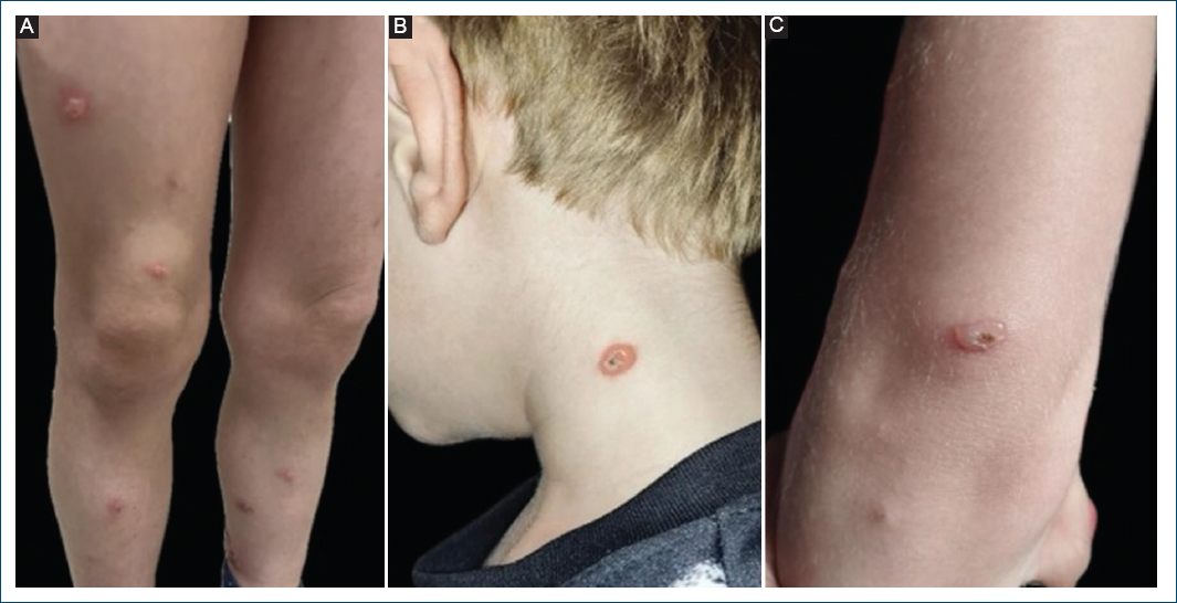
Figure 1. Clinical images. A: multiple tense and grouped vesiculobullous lesions, presenting a “rosette-like” appearance on the lower limbs. B: in detail, lesions arranged in an annular confluence around a vesiconecrotic center on seemingly healthy skin, located in the cervical region. C: vesiculobullous lesion with central ulceration, located on the upper limb.
Based on the characteristic appearance of the lesions, LABD was the leading clinical diagnosis. A skin biopsy, DIF, and laboratory tests before dapsone initiation – the treatment of choice – were requested. Histopathological analysis of perilesional skin from the left leg revealed epidermis with irregular acanthosis, mild spongiosis, and hyperparakeratosis, along with a fibrinoleukocytic crust. The superficial dermis showed a mild perivascular lymphocytic and neutrophilic infiltrate with congestion and edema. DIF demonstrated granular IgA deposits along the BMZ, while IgG, IgM, and C3 were negative (Figs. 2A and B). Salt-split skin testing and in indirect immunofluorescence (IIF) were unavailable. Complete blood count, renal and liver function tests, and glucose-6-phosphate dehydrogenase (G6PD) levels were normal.
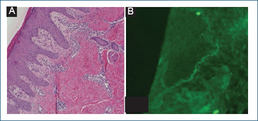
Figure 2. Histopathology and direct immunofluorescence with granular IgA deposition. A: incisional biopsy of a skin lesion on the distal third of the left leg, consistent with a skin fragment showing epidermis with irregular acanthosis, mild spongiosis, and hyperparakeratosis, in addition to an area covered by a fibrinoleucocytic crust. The superficial portion of the dermis shows a mild inflammatory infiltrate composed of lymphocytes and neutrophils, along with congestion and edema. Hematoxylin-eosin, ×10. B: direct immunofluorescence of skin from the distal third of the left leg demonstrating granular deposits of immunoglobulin A in the basement membrane zone. Direct immunofluorescence, ×10.
Due to the granular IgA pattern, the patient was referred to gastroenterology to investigate DH. Serologic testing was negative for anti-endomysial IgA and IgG antibodies and anti-tissue transglutaminase IgA antibodies. In additon, there were no gastrointestinal symptoms, namely on exposure to gluten, which further supported the exclusion of DH.
The differential diagnosis also included bullous impetigo, BP, and epidermolysis bullosa acquisita (EBA). The absence of clinical or laboratory evidence of bacterial infection, poor response to antibiotics, and the typical rosette-like arrangement of tense vesicles on seemingly healthy surrounding skin ruled out bullous impetigo. BP and EBA were considered unlikely given the patient’s age, lesion distribution, and negative IgG and C3 on DIF.
The case was therefore managed as LABD, based on the classic clinical presentation and exclusion of other differentials. Dapsone was initiated at a dose of 0.5 mg/kg/day, but lesions persisted at this dosage. Increasing the dose to 1 mg/kg/day led to full remission within 1 month, despite the absence of a gluten-free diet. The patient remains on maintenance therapy with dapsone 1 mg/kg/day and under quarterly clinical and laboratory follow-up, with no residual or recurrent lesions and no evidence of adverse effects or drug-related toxicity to date.
Discussion
First described by Bowen in 1901,6 LABD is the most frequent autoimmune blistering disease in childhood,4,7 though it remains rare, with an estimated incidence of 0.5-2.3 cases per million individuals per year.4 Blister formation results from an autoimmune response in which IgA-class autoantibodies target antigens in the BMZ of the skin and mucous membranes,1 disrupting dermal-epidermal adhesion and leading to cleavage at multiple levels.5
The primary antigenic targets identified in LABD are the 97-kDa and 120-kDa fragments of BP antigen 2 (BP180/collagen XVII),1,2,4 a transmembrane protein essential for dermal-epidermal adhesion, located in the lamina lucida.4 Although most cases are idiopathic,1,2,4,7 drug-induced LABD – particularly related to vancomycin – has been described in adults.1,4 Genetic predisposition has been associated with HLA types HLA-B8, HLA-DR3, HLA-DQ2, and HLA-Cw7.2,4,7
Clinically, LABD may present with tense, scattered bullae on normal-appearing skin or herpetiform lesions on an erythematous base. Lesions may contain serous or hemorrhagic fluid. In children, the classic “string of pearls” pattern manifests as annular erythematous plaques with peripheral vesicles,2,4 as observed in our patient. This rosette-like arrangement, caused by new vesicle formation at the edges of resolving lesions, is uncommon in adults.1 Pediatric cases typically affect the lower abdomen, thighs, and groin, whereas adult forms more often involve the extensor surfaces, face, and trunk.1 A short prodromal phase with intense pruritus or systemic symptoms such as fever and anorexia may precede the onset of lesions.5
With proper treatment, lesions usually resolve without scarring, although residual pigmentation may persist.5 Mucosal involvement is also common, particularly in the oral and ocular regions.1,4 Oral lesions include painful ulcers, erosive or desquamative gingivitis, and occasionally, scarring.1 Ocular involvement may cause conjunctival hyperemia, ocular discharge, pain, or foreign body sensation; chronic inflammation can result in synechiae or even vision loss.4
Due to its broad clinical spectrum, LABD can clinically mimic other bullous diseases,8 especially DH, a gluten-sensitive dermatosis characterized by pruritic erythematous papules and granular IgA deposition along the BMZ.9 Therefore, definitive diagnosis requires a skin biopsy for hematoxylin-eosin histopathology and DIF.5
Histologically, LABD features a subepidermal blister with a predominantly neutrophilic infiltrate in the papillary dermis,1 though lymphocytes and eosinophils may also be present.2 In DIF, linear IgA deposition along the BMZ is the hallmark finding. However, approximately 20% of patients may display granular deposits.10 Concomitant linear C3 and, less frequently, IgG deposits may be observed, though IgG staining is usually weaker than IgA. When IgG and IgA deposits show similar intensity, distinguishing LABD from BP or other subepidermal autoimmune blistering diseases – such as EBA – becomes challenging.10 In the present case, IgG and C3 deposits were absent.
If a granular IgA pattern is observed, DH should be excluded through negative serologic testing for anti-tissue transglutaminase and anti-endomysial IgA antibodies.3 In uncertain cases, salt-split skin immunofluorescence showing linear IgA deposition on the epidermal side of the split supports LABD.3
Although bullous impetigo, BP, and EBA were initially considered, clinical and immunopathological findings ruled them out. The tense, symmetric, non-fragile vesicles and negative DIF for IgG and C3 were inconsistent with both BP and EBA, while the absence of infection and resistance to antibiotic therapy excluded bullous impetigo.
Spontaneous remission may occur in children within 2-4 years of disease onset,7 pharmacological treatment is usually required. Delayed diagnosis increases the risk of complications, including secondary infection5 or ocular sequelae. Dapsone, a sulfone with immunomodulatory properties, is the first-line treatment. It is typically started at doses above 0.5 mg/kg/day and titrated as needed.7 Most of the patients improve within days; lack of response should prompt diagnosis reassessment. This rapid improvement aligns with recent multicenter data showing marked benefit within the 1st week of dapsone therapy.11
Due to the risk of dapsone-induced hemolysis,5 evaluation of G6PD levels before treatment and regular monitoring of blood counts, bilirubin, lactic dehydrogenase, and aminotransferases are essential.4,7 Cutaneous lesions usually heal without scarring, although mucosal fibrosis may lead to functional impairment.
In DH, by contrast, dapsone provides only symptomatic relief, and strict adherence to a gluten-free diet remains the disease-modifying treatment needed to achieve sustained remission.12
This case describes a patient with typical clinical features of LABD but an atypical granular IgA pattern on DIF, underscoring the importance of comprehensive diagnostic evaluation and exclusion of differentials to ensure optimal outcomes. Despite the non-classical DIF pattern, the presence of rosette-like lesions and negative DH serology supported the clinical diagnosis of LABD. Prompt initiation of dapsone led to complete remission of the lesions despite the absence of a gluten-free diet, highlighting the importance of early recognition and treatment to prevent complications, reduce morbidity, and minimize recurrence in this rare and underrecognized dermatologic condition.
Conclusion
This case demonstrates that linear immunoglobulin A bullous dermatosis (LABD) may present with an atypical granular pattern on direct immunofluorescence, leading to diagnostic uncertainty. Careful clinicopathological correlation and systematic exclusion of differential diagnoses, particularly dermatitis herpetiformis, are essential for establishing the diagnosis. The complete remission achieved with dapsone, even in the absence of a gluten-free diet, reinforces that early recognition of the disease and timely treatment can favorably modify the clinical course, reducing morbidity and preventing complications in pediatric patients.
Funding
None.
Conflicts of interest
None.
Ethical considerations
Protection of human subjects and animals. The authors declare that no experiments on humans or animals were performed for this research.
Confidentiality, informed consent, and ethical approval. The authors have followed their institution’s confidentiality protocols, obtained informed consent from all patients, and secured approval from the Ethics Committee. SAGER guidelines have been followed as applicable to the nature of the study.
Declaration on the use of artificial intelligence. The authors declare that no generative artificial intelligence was used in the writing or creation of the content of this manuscript.
