

Introduction
Multinucleated cell angiohistiocytoma (MCAH) is a rare cutaneous entity of uncertain etiology, characterized by a benign but slowly progressive course.1 First described in 1985 by Smith and Wilson-Jones, it is typically encountered in middle-aged adults, with a female predominance.1 Clinically, MCAH presents as asymptomatic, slowly enlarging reddish-brown papules or nodules, which may occur singly or multiply, usually in a unilateral distribution and often on acral sites, particularly the limbs or dorsal hands.1 However, bilateral or generalized presentations are exceedingly rare.2
Because MCAH is so uncommon, diagnosis is often delayed or mistaken. Its clinical appearance may mimic granuloma annulare, lichen planus, or vascular proliferations such as angiofibroma, dermatofibroma, or Kaposi sarcoma.3
The pathogenesis of MCAH remains controversial. The prevailing hypothesis is that it represents a reactive rather than neoplastic process, a view supported by its benign behavior, absence of extracutaneous spread, lack of malignant transformation, and occasional reports of spontaneous regression.2–4 Some authors have also proposed a hormonal influence, given observed overexpression of estrogen receptor (ER) α in some cases, which might explain the female predominance; however, ER positivity has not been consistently reproduced across all reported cases.2,3 Alternative views include considering MCAH a variant of dermatofibroma or associating it with an altered immune status.4 Interestingly, Ogawa et al., reported a case suggesting the possible onset of MCAH after Treponema pallidum infection.4
Recent non-invasive imaging tools, such as dermoscopy and reflectance confocal microscopy, have been reported to aid in the clinical recognition of MCAH by improving the correlation between clinical and histopathologic findings.5 These techniques may improve clinical recognition and diagnostic accuracy, particularly in atypical or generalized presentations.
Histopathologically, MCAH typically shows, in the superficial dermis, proliferation of small vessels embedded in a dense collagenous stroma composed of interstitial fibrohistiocytic cells and bizarre multinucleated giant cells with angular or scalloped outlines. These multinucleated cells, while characteristic, are not pathognomonic.6,7 Immunohistochemistry, although not indispensable, can support the diagnosis: The multinucleated giant cells often express vimentin, factor XIIIa, and CD68, and are typically negative for factor VIII, CD34, and S100.7 Some studies have also observed a positive correlation between endothelial CD68 expression and lesion size, and an inverse correlation between multinucleate cell CD68 expression and the occurrence of multiple lesions.4
The rarity of the condition and its nonspecific presentation make diagnosis challenging, often requiring histopathological confirmation.
Therapeutic interventions are primarily for cosmetic or symptomatic reasons. Reported options include topical or intralesional corticosteroids, surgical excision, cryotherapy, argon laser, intense pulsed light, CO2 laser, and in one case, it was described the use of potassium-titanyl-phosphate laser combined with intralesional corticosteroids.4,8 Due to heterogeneity and scarcity of data, there is no consensus on optimal management.
To date, approximately 150 cases of MCAH have been reported in the literature, with only a small fraction representing generalized variants; it is likely underrecognized, due to both its indolent nature and the possibility of misdiagnosis.2,4 Given the limited experience with MCAH, case reports contribute meaningfully to expanding knowledge regarding clinical variants, diagnostic pitfalls, and therapeutic outcomes. In this work, we present a new clinical case of MCAH, discuss its diagnostic challenges, and place it in the context of the existing literature.
Case report
A 50-year-old male, ex-smoker, with a medical history of typhoid fever, malaria, Bell’s palsy, steatohepatitis, and dyslipidemia, presented with multiple erythematous-violaceous papules and plaques, ranging from 3 to 12 mm in diameter, distributed over the dorsal aspect of the left hand (Fig. 1). The lesions were asymptomatic but showed a slow, progressive enlargement over several months, with some evolving into firm subcutaneous nodules. There was no history of local trauma to the left upper limb before the appearance of the lesions.
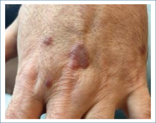
Figure 1. Clinical presentation with violaceous papules on the dorsum of the left hand.
A punch biopsy demonstrated a dermal proliferation of small vessels within a collagenous stroma containing multinucleated giant cells, consistent with MCAH. Considering the benign course of the disease and the absence of functional or aesthetic impairment, a conservative management strategy with active clinical surveillance was adopted.
Clinical photographs were obtained in September 2024 (Fig. 1) and again in December 2025 (Fig. 2), demonstrating no significant changes in the size, number, or morphology of the lesions during a follow-up period of approximately 15 months, supporting the decision for continued active surveillance.
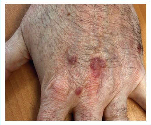
Figure 2. Follow-up clinical image obtained in December 2025, showing no significant changes in the lesions.
Discussion and conclusion
This case underscores the importance of including MCAH in the differential diagnosis of vascular and fibrohistiocytic skin lesions. Clinically, MCAH may resemble granuloma annulare, lichen planus, dermatofibroma, microvenular hemangioma, or Kaposi sarcoma, given its erythematous-violaceous coloration and papulonodular morphology so histopathology remains essential for diagnosis, showing dermal vascular proliferation within a collagenous stroma and characteristic multinucleated giant cells, distinguishing it from other mimickers.
Although benign and indolent, MCAH is often underrecognized, potentially leading to unnecessary biopsies or excisions. Accurate diagnosis, therefore, relies on clinicopathologic correlation and awareness of this rare entity. By reporting this case, we aim to contribute to the growing body of literature on MCAH and to reinforce the value of multidisciplinary collaboration in the assessment of uncommon cutaneous tumors.
Funding
None.
Conflicts of interest
None.
Ethical considerations
Protection of human subjects and animals. The authors declare that no experiments on humans or animals were performed for this research.
Confidentiality, informed consent, and ethical approval. The authors have followed their institution’s confidentiality protocols, obtained informed consent from all patients, and secured approval from the Ethics Committee. SAGER guidelines have been followed as applicable to the nature of the study.
Declaration on the use of artificial intelligence. The authors declare that no generative artificial intelligence was used in the writing or creation of the content of this manuscript.
