

Introduction
Mycetoma is a chronic, destructive granulomatous infection that primarily affects the skin and subcutaneous tissues, with frequent extension to fascia, muscle, and bone.1 It is clinically characterized by progressive painless swelling, multiple fistulous tracts, and the pathognomonic drainage of pus containing colored grains.2 Mycetoma is classified as eumycetoma (fungal) or actinomycetoma (bacterial) depending on the causative organism.3 It is endemic in the “mycetoma belt” (tropical and subtropical regions between 30°N and 15°S), including Sudan, India, and parts of Latin America, with sporadic cases elsewhere.4 It typically affects body parts that are frequently exposed and in direct contact with soil, most commonly the feet, followed by the lower limbs and hands.1 The usual route of infection is traumatic inoculation of the causative organism, often through thorn pricks, splinters, or minor, unnoticed wounds.5 Mycetoma progresses slowly but relentlessly, commonly causing delayed diagnosis and resulting in profound disability, disfigurement, and social stigma, leading to isolation.6 Early recognition of the causative agent and prompt medical-surgical treatment are essential to prevent these outcomes.4
Case report
A 34-year-old Moroccan man presented to our Dermatology department with a 2-year history of painless swelling of the right foot studded with multiple sinuses. The lesion had appeared after a wooden splinter injury sustained while working barefoot in the fields. He had no comorbidities or immunosuppression. Clinical examination revealed a large ulcerated plaque with a firm, indurated swelling that fistulized with discharge of seropurulent fluid with pale grains, located on the heel and sole of the left foot (Fig. 1). Computed tomography (CT) scan showed ill-defined lytic and sclerotic lesions in the posteroinferior calcaneus, along with soft-tissue infiltration adjacent to the Achilles tendon (Fig. 2). Magnetic resonance imaging (MRI) revealed the characteristic ‘dot-in-circle’ sign as well as multiple fistulous tracts involving the calcaneus. Mycetoma was strongly suspected based on the clinical and radiological findings. The patient underwent surgery. Histopathological examination showed an extensively ulcerated epidermis replaced by a hyperplastic, fleshy growth composed of radial capillaries and intermingled with an inflammatory infiltrate rich in neutrophils. The dermis and hypodermis were separated by a suppurative inflammatory infiltrate rich in altered neutrophils surrounding clusters of bacteria composed of strongly basophilic grains and filaments. This infiltrate extended to and dissociated the underlying bone tissue, which showed dystrophic and ragged bone trabeculae; no signs of malignancy were present (Fig. 3). Gram staining showed many Gram-positive filamentous branching bacilli, and culture identified actinomycetoma (Actinomadura spp.).
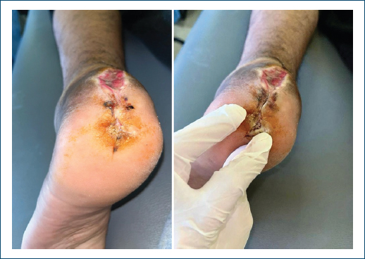
Figure 1. Fistulized subcutaneous swelling of the sole of the left foot with whitish granular discharge.
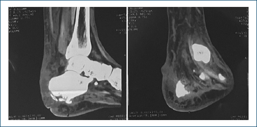
Figure 2. Sagittal and coronal reformats of computed tomography of ankle show multiple osteolytic areas in calcaneum with periosteal reaction at the lower end of tibia and fibula with soft-tissue involvement, respectively.
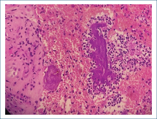
Figure 3. Microphotograph showing Actinomadura grains surrounded by multi-inflammatory cells (H&E staining, 200×).
He received prolonged antibiotic therapy for 12 months, consisting of amoxicillin-clavulanate (30 mg/kg/day), amikacin (15 mg/kg/day), and oral trimethoprim-sulfamethoxazole (TMP-SMX) (40 mg/kg/day). Local wound care was provided daily throughout the treatment period and continued until complete healing. It consisted of gentle cleansing with normal saline to irrigate sinuses and remove debris and seropurulent discharge, followed by application of silver sulfadiazine cream for its broad-spectrum antimicrobial activity and to reduce secondary bacterial colonization in the chronic fistulizing wounds. The treated areas were then covered with sterile compresses and bandaging to maintain a moist healing environment, facilitate drainage, protect the site from further trauma, and promote granulation tissue formation. At the 12-month follow-up, complete healing of ulcers and marked clinical improvement were observed, with no recurrence despite initial bone involvement.
Discussion
Mycetoma was first described as a distinct clinical entity in 1842 by Gill in Madurai, India, where it was termed “Madura foot”.7 The term “mycetoma” was later introduced by Carter in 1860 to describe the tumor-like swelling with characteristic grains.8 The disease is caused by over 56 microorganisms and is classified into actinomycetoma (bacterial) and eumycetoma (fungal).9 Actinomycetoma, often caused by aerobic Gram-positive bacteria such as Nocardia, Streptomyces, and Actinomadura species, tends to be more aggressive and disseminative,3 while eumycetoma is predominantly due to Madurella mycetomatis,5 a dematiaceous fungus.10 It mainly affects young men aged 20–40 years, most commonly farmers or field workers who sustain minor, often unnoticed injuries while walking barefoot or working in soil contaminated by the causative organisms.11 It is endemic in tropical and subtropical regions, including countries such as Sudan, Somalia, Senegal, Yemen, India, Mexico, and Venezuela, although sporadic cases have been reported in temperate climates.12 In May 2016, the World Health Organization recognized mycetoma as a neglected tropical disease due to its high prevalence among impoverished and remote rural populations in developing countries.7 Clinically, mycetoma starts as painless subcutaneous swelling, which frequently contributes to significant diagnostic delay.1 Pain usually appears only later, secondary to superimposed bacterial infection or extension to underlying bone.13 Following traumatic inoculation, the causative organisms aggregate and surround themselves with a dense, cement-like matrix, forming characteristic grains. This protective structure shields the pathogen from both the host immune response and antimicrobial agents, thereby explaining the chronicity and therapeutic resistance commonly observed.13 The lower limbs, particularly the foot, are by far the most commonly affected sites, followed by the hand.5 Less frequent locations include the arm, buttocks, shoulder, trunk, and neck.7 The interval between initial inoculation and definitive diagnosis is highly variable, ranging from a few months to as long as 60 years,14 as seen in our patient, who was diagnosed after a 2-year history of progressive symptoms. Mycetoma can mimic a wide range of infectious and non-infectious conditions, including cutaneous tuberculosis, schistosomiasis, leishmaniasis, botryomycosis, chromoblastomycosis, sporotrichosis, as well as non-infectious disorders such as benign soft-tissue tumors, Kaposi sarcoma, and skin malignancies.2,13 The differential diagnosis is therefore extensive, particularly in non-endemic areas. Definitive diagnosis relies on the combination of characteristic clinical features, notably the classic triad of painless tumefaction, multiple draining sinuses, and seropurulent discharge containing grains, histopathology, and culture revealing grains of fungal hyphae or bacteria within a granulomatous reaction.2 Although the diagnosis can often be strongly suspected on clinical grounds alone, it is not conclusive. Accurate and specific diagnostic tools are therefore required to confirm the etiology and guide appropriate treatment.14 Characteristic imaging findings play a key role: MRI typically reveals the “dot-in-circle” sign, consisting of tiny hypointense foci (representing grains) within hyperintense spherical lesions on T2-weighted and short TI inversion recovery (STIR) sequences;4 CT effectively delineates bone involvement and assesses the overall extent of disease.13 Additional supportive methods include fine-needle aspiration cytology and, increasingly, molecular techniques such as PCR and sequencing to precisely identify the causative organism.14 Management of actinomycetoma involves surgical excision combined with prolonged antibiotic therapy; it generally responds more rapidly and favorably to medical treatment than eumycetoma.2 Trimethoprim-TMP-SMX remains the cornerstone and gold standard treatment. To improve cure rates and shorten treatment duration, combination regimens that include amikacin, carbapenems, or amoxicillin-clavulanic acid are frequently used and widely recommended.15 In our patient, a 34-year-old Moroccan man with a typical actinomycetoma due to Actinomadura spp. Following a splinter injury, early combined medical-surgical treatment led to complete ulcer healing and marked clinical improvement with no recurrence at 12 months despite initial bone involvement. Although Morocco is not considered part of the classic mycetoma belt, the potential for sporadic cases linked to agricultural work and barefoot exposure highlights the need for increased awareness among dermatologists in North Africa. Prolonged follow-up is essential, given the high risk of late relapse.
Conclusion
This case reinforces the importance of including mycetoma in the differential diagnosis of chronic foot lesions, even in countries outside the classic “mycetoma belt.” In settings such as Morocco, greater clinician awareness combined with systematic use of accessible diagnostic tools (histology and culture) can significantly reduce diagnostic delays. Ultimately, the most effective approach to preventing severe disability and amputation remains primary prevention through adequate foot protection in rural populations, together with early combined medical-surgical intervention to preserve limb function.
Funding
None.
Conflicts of interest
None.
Ethical considerations
Protection of human subjects and animals. The authors declare that no experiments on humans or animals were performed for this research.
Confidentiality, informed consent, and ethical approval. The authors have followed their institution’s confidentiality protocols, obtained informed consent from all patients, and secured approval from the Ethics Committee. SAGER guidelines have been followed as applicable to the nature of the study.
Declaration on the use of artificial intelligence. The authors declare that no generative artificial intelligence was used in the writing or creation of the content of this manuscript.
