

Introduction
Rosacea and cutaneous lupus erythematosus (CLE) are inflammatory dermatoses that may share similar clinical features, such as persistent facial erythema and inflammatory papules and plaques, complicating the differential diagnosis.1 The potential coexistence of both conditions further contributes to this diagnostic challenge, requiring a careful and nuanced approach to avoid inappropriate or unnecessary therapeutic interventions.2 The objective of this report is to underscore the importance of an accurate differential diagnosis of new-onset skin lesions in patients with systemic lupus erythematosus (SLE), particularly in distinguishing CLE from other inflammatory dermatoses such as rosacea.
Case report
We present a 47-year-old woman diagnosed with SLE in 2003, with cutaneous, articular, and hematological involvement. She was under treatment with belimumab 200 mg/week, hydroxychloroquine 400 mg/day, and methotrexate 10 mg/week, with apparent clinical control of the disease. The patient developed a facial dermatosis over approximately 5 months, initially suspected to represent CLE, which led to consideration of initiating anifrolumab. She was referred to the Dermatology clinic for diagnostic confirmation and possible reassessment of her systemic treatment. On examination, multiple erythematous papules and plaques with a hyperkeratotic appearance, associated with pustules on the frontal, periorbital, and malar regions, were observed, with sparing of the nasolabial fold (Fig. 1). Laboratory tests showed no anemia, thrombocytopenia, renal dysfunction, or proteinuria. Anti-double-stranded DNA antibodies and complement were within normal limits. As the clinical and laboratory findings were not suggestive of active lupus, a skin biopsy was performed. Histology revealed a moderate perivascular and periadnexal dermal inflammatory infiltrate, with non-necrotizing epithelioid granulomas and a subepidermal vesicle/pustule. The epidermis showed irregular acanthosis, mild spongiosis, and hyperkeratosis with intracorneal neutrophils (Fig. 2). Based on the clinical and histopathological findings, a diagnosis of papulopustular rosacea was established. Treatment was initiated with isotretinoin 20 mg/day in combination with prednisolone 20 mg/day, followed by gradual tapering of the corticosteroid. As clinical improvement was observed, the isotretinoin dose was progressively titrated up to 40 mg/day, allowing for further reduction and discontinuation of systemic corticosteroid therapy. Follow-up assessment demonstrated marked clinical improvement with complete resolution of the lesions (Fig. 3). Following exclusion of cutaneous lupus, the patient continued her previously established immunomodulatory therapy. At the most recent follow-up, 15 months after treatment initiation, the patient had been off isotretinoin for 8 months and remained in complete clinical remission.
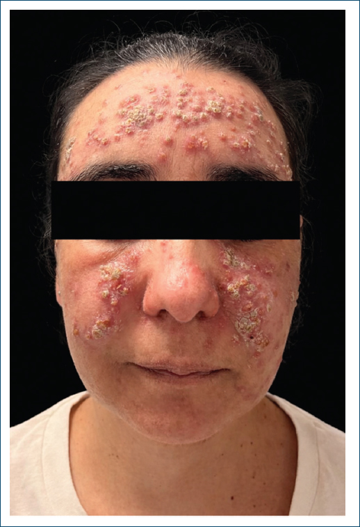
Figure 1. Facial dermatosis in a patient with longstanding systemic lupus erythematosus, showing erythematous papules, plaques, and pustules involving the frontal, periorbital, and malar regions, with characteristic sparing of the nasolabial folds.
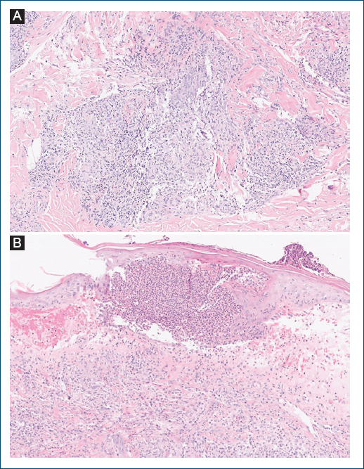
Figure 2. Histopathological examination showing a moderate perivascular and periadnexal inflammatory infiltrate in the dermis, with non-necrotizing epithelioid granulomas and an associated subepidermal vesicle/pustule (A). The epidermis exhibits irregular acanthosis, mild spongiosis, and hyperkeratosis with intracorneal neutrophils (B).
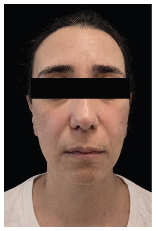
Figure 3. Complete clinical resolution of the facial eruption following treatment with isotretinoin and a short course of systemic corticosteroids.
Discussion
In patients with SLE, the appearance of new cutaneous lesions raises concern for CLE, given the high prevalence of skin involvement and its potential association with systemic disease activity. However, CLE encompasses a heterogeneous spectrum of subtypes, including acute, subacute, and chronic forms, each with distinct clinical morphologies, distributions, and prognostic implications.3 This variability, combined with the frequent facial involvement of lupus, may complicate the clinical assessment when other inflammatory dermatoses coexist.
Rosacea represents a diagnostic challenge in patients with SLE due to its predilection for the centrofacial region and potential clinical overlap with CLE. Rosacea is typically characterized by persistent centrofacial erythema, episodic flushing, telangiectasia, and inflammatory papules or pustules. Although ultraviolet exposure may exacerbate rosacea, lesions generally lack the characteristic photodistribution and scarring seen in certain CLE subtypes. In contrast, CLE comprises a heterogeneous spectrum of clinical presentations. Acute and subacute forms usually present as non-scarring, photosensitive erythematous lesions, whereas chronic CLE, particularly discoid lupus erythematosus, manifests as well-defined plaques with scale, follicular plugging, dyspigmentation, and permanent scarring.4 Histopathology plays a crucial role when clinical distinction is uncertain. CLE is characterized by interface dermatitis with basal vacuolar change, apoptotic keratinocytes, thickening of the basement membrane, and a superficial and deep perivascular and periadnexal lymphocytic infiltrate, frequently accompanied by dermal mucin deposition. Conversely, rosacea lacks interface changes and typically demonstrates perifollicular and perivascular lymphohistiocytic inflammation, dilated superficial vessels, dermal edema, and, in some cases, granulomatous inflammation.1 In this case, although the initial clinical presentation raised suspicion for lupus-related cutaneous involvement, histological and immunopathological findings were not supportive of CLE, allowing this diagnosis to be confidently excluded. Recognition of rosacea as the underlying condition had important therapeutic implications, permitting continuation of the existing immunomodulatory regimen without unnecessary escalation.
Conclusion
Overall, this case highlights the importance of maintaining a broad differential diagnosis when evaluating new cutaneous findings in SLE patients. Accurate distinction between CLE and mimicking inflammatory dermatoses relies on careful clinicopathological correlation and assessment of systemic disease activity, ensuring appropriate treatment decisions and optimized patient care.
Funding
None.
Conflicts of interest
None.
Ethical considerations
Protection of human subjects and animals. The authors declare that no experiments on humans or animals were performed for this research.
Confidentiality, informed consent, and ethical approval. The authors have followed their institution’s confidentiality protocols, obtained informed consent from all patients, and secured approval from the Ethics Committee. SAGER guidelines have been followed as applicable to the nature of the study.
Declaration on the use of artificial intelligence. The authors declare that ChatGPT was used for grammatical correction.
