
Introduction
Pilomatricoma, or calcifying epithelioma of Malherbe, is an uncommon benign adnexal tumor derived from the hair matrix, accounting for about 0.12% of all cutaneous neoplasms. It occurs most commonly in children and young adults and shows a slight predominance in females.1 Clinically, it is usually a slowly enlarging, well-defined lesion that appears as a cystic or firm dermal nodule measuring approximately 0.5–2 cm. The most frequent sites of involvement are the head and neck, followed by the upper limbs and trunk.2 Although the classic form is small and discreet, unusual variants such as bullous, ulcerated, anetodermic, lymphangiectatic, and giant pilomatricoma have also been described. Bullous pilomatricoma is a rare variant that is frequently misdiagnosed, with only a limited number of cases documented in the literature.3 The underlying mechanism responsible for its characteristic bullous appearance remains uncertain. Although trauma has been proposed as a potential contributing factor, a definitive causal relationship has not yet been demonstrated.
We report a case of bullous pilomatricoma presenting as a gradually enlarging mass over the lower back in a young male, initially misinterpreted as a malignant tumor in cytology, emphasizing the importance of clinicopathologic correlation in such cases.
Case report
A 23-year-old male presented with a complaint of swelling over the right lower back region for the past 2 months. The mass had gradually increased in size and caused mild discomfort when lying down. There was no history of trauma, discharge, ulceration, or systemic symptoms. The patient’s medical and family history were unremarkable.
On local examination, a soft, mobile, tender, subcutaneous nodule with overlying bulla measuring approximately 3 × 2.5 cm was noted on the right lower back. The surrounding skin showed erythema. The swelling was mobile but adherent to the overlying skin. No regional lymphadenopathy was detected.
Fine-needle aspiration cytology revealed loosely cohesive clusters and singly scattered atypical cells in a blood-mixed lipidic background. Individual tumor cells were round to oval, showing moderate pleomorphism, a scant to moderate amount of cytoplasm, round to oval nuclei showing coarse chromatin and inconspicuous nucleoli. Occasional giant cells were also noted along with a few stromal fragments. Cytological impression was suggestive of malignancy, and excision biopsy was advised (Fig. 1A).
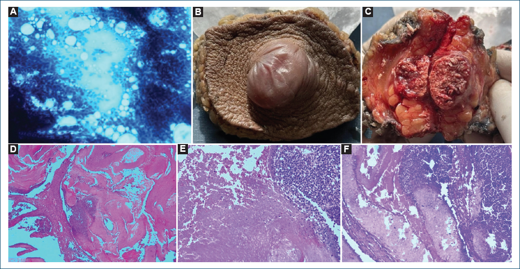
Figure 1. A: FNA smear showing loosely cohesive clusters with singly scattered atypical cells in a blood mixed lipidic background. B: gross picture showing a bullous nodule measuring 3 Å~ 2.5 cm in size. C: cut surface of bullous nodule showing a well-circumscribed, gray-tan mass. D-F: biopsy of bullous nodules showing lobules of small basaloid cells at the periphery with underlying ghost cells toward the center (H&E).
We received a skin-covered tissue measuring 8 × 6 × 3 cm with a bulla over the skin measuring 3 cm in diameter. The cut surface showed a well-circumscribed, gray tan mass measuring 2 × 1.8 × 1.5 cm, which was 1 cm away from the deep resected margin and almost abutting the overlying skin (Fig. 1B and C).
Microscopic examination showed a well-circumscribed dermal tumor with an overlying bulla composed of lobules of basaloid cells at the periphery transitioning into anucleate eosinophilic “ghost” cells toward the center. Foci of calcification and foreign-body giant cell reactions were seen. No significant mitotic activity, necrosis, or vascular invasion was noted. The features were consistent with bullous pilomatricoma (Fig. 1D, Fig. 1E, F).
Discussion
Pilomatricoma is a benign tumor arising from hair matrix cells. This tumor, originally thought to arise from sebaceous glands, was first described by Malherbe and Chenantais in 1880, who named it “calcifying epithelioma.” Later, in 1961, Forbis and Helwig introduced the term “pilomatrixoma” in their publication to better reflect its histological origin from the hair matrix.4,5
Clinically, pilomatricoma presents as a deeply situated, firm subcutaneous mass fixed to the overlying skin, usually measuring 0.5–3 cm in diameter, and only rarely reaching up to 5 cm.6 Although benign, its clinical and cytological features can sometimes simulate malignancy. The most common sites are the head, neck, and upper extremities. Occurrence on the trunk or back, as in this case, is distinctly rare.
Histopathologically, pilomatricoma shows a biphasic pattern – basaloid cells and ghost (shadow) cells with calcification, sometimes accompanied by foreign-body giant cell reaction. These features differentiate it from malignant pilomatrix carcinoma, which shows infiltrative growth, nuclear pleomorphism, frequent mitoses, and necrosis. Several case reports have described unusual presentations of pilomatricoma at sites such as the chest wall and sternum, mimicking malignancy both clinically and cytologically (Table 1).7,8
Table 1. Cases of pilomatrioma misdiagnosed on cytology/clinically
| Year | Author/Ref. | Site | Cytological diagnosis | Clinical diagnosis |
|---|---|---|---|---|
| 1997 | Inui et al.9 | Right thigh | – | Malignancy |
| 2004 | Yuca et al.2 | Left preauricular region | Non-contributory | – |
| 2018 | Harsha et al.8 | Sternum | Malignancy | – |
| 2020 | Sabater et al.1 | Left parotid region | – | Cutaneous squamous cell carcinoma |
| 2023 | Birman et al.4 | Right forearm | – | Malignancy |
| 2023 | Bhole et al.7 | Chest | Round cell neoplasm | – |
| 2025 | Current case | Right lower back | Malignancy |
The bullous variant of pilomatricoma represents only about 2% of all reported pilomatricoma cases.10 Two main hypotheses have been proposed to explain the development of bullous pilomatricoma. The more widely accepted theory suggests that mechanical stimulation acts as a trigger for the blister-like change. In this model, the tumor compresses adjacent lymphatic vessels, resulting in lymphatic dilatation, lymph stagnation, and chronic inflammatory cell infiltration, ultimately producing dermal edema around the lesion9. Motegi et al. further proposed that mechanical stress or bacterial infection may induce cytokines and chemokines, leading to the recruitment of matrix metalloproteinase (MMP)-positive macrophages.11
The possibility of pilomatricoma should always be considered when evaluating cytological smears from any subcutaneous swelling to prevent misdiagnosis. Such atypical presentations underline the importance of histopathological confirmation. Complete surgical excision with clear margins remains the treatment of choice. Recurrence is uncommon and generally attributed to incomplete excision.
Conclusion
Bullous pilomatricoma should be considered in the differential diagnosis of firm, bullous, subcutaneous masses, even in atypical locations such as the back or trunk. Awareness of its cytological spectrum can prevent misdiagnosis as a malignant tumor. Histopathological examination remains the gold standard for diagnosis, and complete excision ensures an excellent prognosis.
Funding
None.
Conflicts of interest
None.
Ethical considerations
Protection of human subjects and animals. The authors declare that no experiments on humans or animals were performed for this research.
Confidentiality, informed consent, and ethical approval. The authors have followed their institution’s confidentiality protocols, obtained informed consent from all patients, and secured approval from the Ethics Committee. SAGER guidelines have been followed as applicable to the nature of the study.
Declaration on the use of artificial intelligence. The authors declare that no generative artificial intelligence was used in the writing or creation of the content of this manuscript.
