

Introduction
Pseudoepitheliomatous keratotic and micaceous balanitis (PKMB) is an exceedingly rare, non-venereal dermatosis of the glans penis, first described by Lortat-Jacob and Civatte in 1961.1 It has typically been reported in elderly men, frequently those circumcised later in life, and clinically presents as one or more thickened hyperkeratotic plaques with adherent, silvery, mica-like scales on the glans.2
The clinical course of PKMB is slow and often asymptomatic, although patients may report irritation, pruritus, dysuria, or other urinary symptoms.3,4 Histologically, PKMB is characterized by acanthosis, pronounced hyperkeratosis, and pseudoepitheliomatous hyperplasia, typically lacking cytological atypia. However, dysplastic changes may be present in a subset of cases.5,6
The etiology remains unclear but PKMB is often associated with acquired phimosis and Lichen Sclerosus (LS). Given that acquired phimosis in adults is almost always secondary to LS, it’s plausible that PKMB and LS represent different expressions within the same pathological spectrum.7
The prognosis of PKMB varies depending on the severity and extent of the lesions. Historically considered benign, PKMB is now widely viewed as a lesion with malignant potential. Increasing evidence suggests that PKMB may evolve through distinct stages – from a plaque phase to verrucous carcinoma and, in some cases, invasive squamous cell carcinoma.2,8 Early recognition and appropriate treatment are essential to minimize the risk of malignant transformation.5
Herein, we report a case of PKMB in an adolescent male, successfully managed with surgical excision. To our knowledge, he is the youngest patient among the fewer than 50 cases reported in the literature to date.
Case report
A 17-year-old Caucasian male presented with a 1-year history of an asymptomatic white lesion on the penis. He had no relevant past medical history and had not yet initiated sexual activity.
On examination, a well-demarcated, white plaque measuring 15 × 10 mm was observed on the balanopreputial sulcus (Fig. 1). It was mobile over the underlying tissues and had a firm, callous-like consistency.
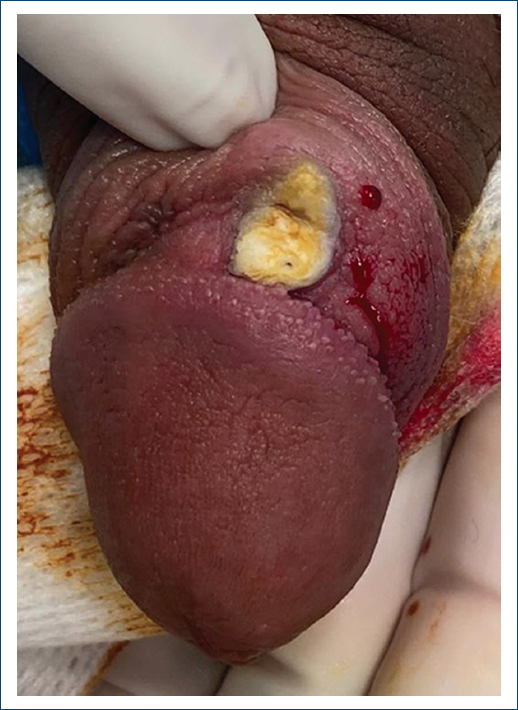
Figure 1. Well-demarcated, white, keratotic plaque on the balanopreputial sulcus of an uncircumcised adolescent, before surgical excision. Note: the yellow coloration is due to the antiseptic povidone-iodine.
The primary diagnostic hypothesis was PKMB, and an excisional biopsy was performed. Histopathological analysis showed acanthosis, hyperkeratosis with parakeratosis, and epithelial hyperplasia with a focal pseudoepitheliomatous appearance, confirming the clinical suspicion (Figs. 2 and 3).
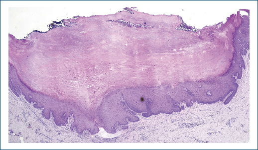
Figure 2. H&E ×10. Well-demarcated lesion showing irregular acanthosis and epithelial hyperplasia, with a central depression filled with compact keratin. Surgical margins are free of disease.
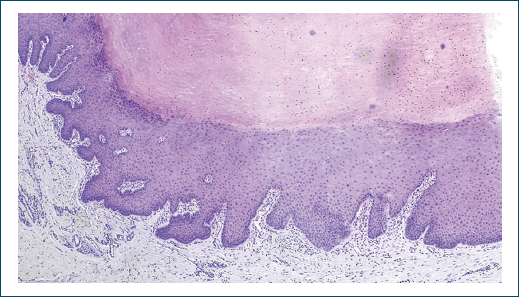
Figure 3. H&E × 40. Parakeratotic hyperkeratosis and acanthosis with focal pseudoepitheliomatous hyperplasia.
Routine laboratory tests, including serology for HIV, syphilis, and hepatitis B and C, were within normal limits or negative.
Two months after surgical excision, follow-up revealed a slightly indurated scar consistent with fibrosis. An intralesional injection of triamcinolone acetonide (20 mg/mL) was administered, with subsequent improvement. The patient has now been followed for approximately 2 years, with no evidence of recurrence (Fig. 4).
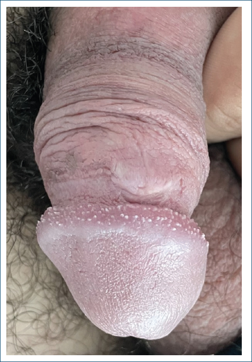
Figure 4. Twenty-one months after surgical excision, with no evidence of recurrence.
Discussion
PKMB is a rare, chronic, non-venereal dermatosis of the glans penis, most reported in elderly males.
Our case stands out due to the patient’s young age – 17 years old – well below the typical age reported in the literature, where most presentations occur after the sixth decade of life.4 This atypical presentation expands the known age spectrum of this disease and highlights that, though rare, it must be considered in the differential diagnosis of keratotic penile lesions in young patients.
The diagnosis of PKMB is based on clinicopathological correlation. Clinically, it presents as verrucous or hyperkeratotic plaques with adherent, silvery, mica-like scales. The differential diagnosis is broad and includes hypertrophic lichen planus, LS, verrucous carcinoma, and squamous cell carcinoma in situ.9 Therefore, histopathological examination is essential to establish a definitive diagnosis. In our patient, the biopsy revealed the typical features of PKMB, namely, acanthosis, compact hyperkeratosis with parakeratosis, and pseudoepitheliomatous hyperplasia without dysplasia, confirming the benign nature of the lesion at evaluation.6,9 However, it is essential to reiterate that PKMB is a condition with malignant potential. Its progression to verrucous carcinoma or invasive SCC is well documented, with variable rates of malignant transformation, necessitating timely diagnosis and appropriate treatment.1,7
Although the etiology remains unclear, it has been hypothesized that PKMB may represent a pseudoepitheliomatous response to chronic irritation or inflammation, possibly with a lichenoid background or a variant of LS.4,7 In our patient, neither condition was identified, further underscoring the atypical presentation.
Therapeutic approaches are not standardized due to the condition’s rarity.10 Reported treatments include topical corticosteroids, topical 5-fluorouracil, cryotherapy, carbon dioxide laser ablation, and complete surgical excision. Surgical excision remains the most definitive treatment, as it allows full histopathological assessment to exclude malignancy and is often curative.9,11 Although there are no standardized treatment guidelines for PKMB, management should be tailored to the clinical presentation and histopathological findings. The choice of therapy depends on the severity and extent of the lesion, as well as the presence or absence of cytological atypia or features suggestive of malignant transformation. In our case, complete surgical excision was curative, with no recurrence observed after 2 years of follow-up.
This case documents an uncommon case of PKMB in an adolescent, expanding the known age spectrum, reinforces the importance of early biopsy and diagnosis, and documents successful surgical management without recurrence.
Conclusion
Pseudoepitheliomatous keratotic and micaceous balanitis is an exceptionally rare penile dermatosis, particularly in adolescents. This case underscores the importance of clinicopathological correlation for accurate diagnosis. Given its recognised malignant potential, early biopsy and appropriate treatment are essential. Careful long-term follow-up is recommended to promptly detect recurrence or malignant transformation.
Funding
None.
Conflicts of interest
None.
Ethical considerations
Protection of human subjects and animals. The authors declare that no experiments on humans or animals were performed for this research.
Confidentiality, informed consent, and ethical approval. The authors have followed their institution’s confidentiality protocols, obtained informed consent from all patients, and secured approval from the Ethics Committee. SAGER guidelines have been followed as applicable to the nature of the study.
Declaration on the use of artificial intelligence. The authors declare that no generative artificial intelligence was used in the writing or creation of the content of this manuscript.
