

Introduction
Syringomas are benign adnexal neoplasms characterized by distinctive ductal differentiation. They have a higher incidence in females and in Asian populations and have been associated with certain genetic syndromes (such as Down syndrome) and metabolic conditions, including diabetes mellitus. They typically present as multiple, asymptomatic, well-defined, skin-colored papules and generally do not require treatment unless for cosmetic concerns.1 The etiology of syringomas remains controversial, with evidence supporting roles for both genetic predisposition and hormonal imbalances.2 The risk of malignant transformation is negligible, and treatment is generally not required.1
Williams syndrome (WS) is a rare, multisystemic genetic disorder caused by contiguous gene deletions on chromosome 7,3 including genes implicated – or possibly implicated – in cutaneous and connective-tissue features, such as ELN (elastin), CLDN3 (claudin-3), CLDN4 (claudin-4), LIMK1, and NCF1, among others. Consequently, WS may affect the skin, leading to softer skin, premature graying of the hair, increased wrinkling, and abnormal scarring.4
To date, an association between syringomas and WS has not been reported in the literature.
Case report
A 24-year-old male patient with WS presented to the Dermatology Clinic with multiple, asymptomatic, millimetric, monomorphic, erythematous-yellowish papular lesions on the chest, present since the age of five (Fig. 1). No triggering or aggravating factors were identified, and no other cutaneous abnormalities were noted. Dermoscopy revealed multiple rounded, erythematous-orange lesions on a homogeneous pink background, along with fine, subtle linear vessels on the surface, without a clear arborizing or glomerular pattern, or a pigment network (Fig. 2). There was no family history of similar cutaneous lesions.
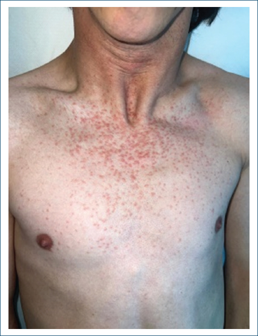
Figure 1. Clinical photograph of multiple eruptive syringomas in the upper thoracic region.
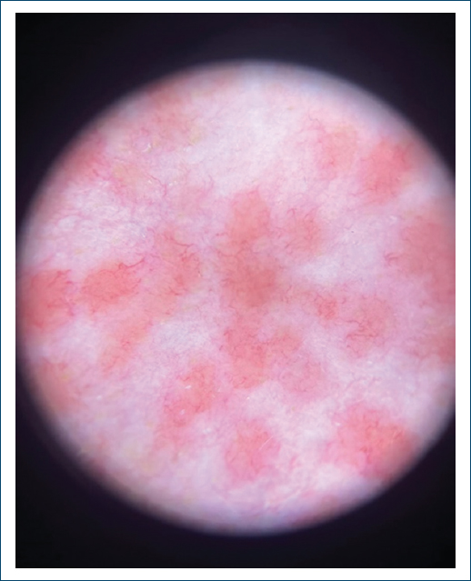
Figure 2. Dermoscopy showing erythematous-orange lesions on a homogeneous pink background, along with fine, subtle linear vessels on the surface.
A clinical hypothesis of syringomas or another type of adnexal tumor was considered, and an incisional biopsy was performed. Histopathological examination revealed superficial and mid-dermal ductal structures lined by a double layer of epithelial cells, with lumina either optically empty or filled with eosinophilic material – findings consistent with syringomas (Fig. 3).
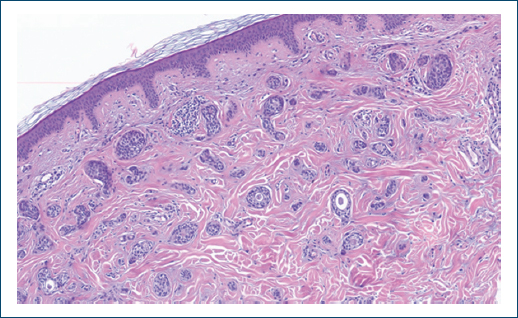
Figure 3. Histopathological examination showing dermal ductal structures, consistent with syringomas.
The available therapeutic options were explained to the patient and his family; however, they opted against treatment given the benign nature of the lesions and the absence of functional or esthetic impact.
Discussion
We report a case of thoracic syringomas in a patient with WS. Although syringomas are more commonly found in the periorbital region, they can also appear in other locations, such as the thoracic and cervical regions, as observed in our case.1
The definitive diagnosis of syringomas relies on histopathology, which typically reveals multiple nests of cells with central ductal differentiation forming lumina lined by an eosinophilic cuticle, within a sclerotic stroma, confined to the superficial dermis, similar to our findings.1 However, the typical clinical presentation, along with dermoscopic findings, is clinically relevant.2 Although dermoscopic features are not yet well-defined in the literature, our observations were consistent with previously reported features, namely pinpoint and small linear vessels on a diffuse orange-pink background. Notably, white globules or pigment networks, which have also been described, were not observed in this case.5–7
A potential association with WS has not been established. Nevertheless, other genetic syndromes have been linked to this condition, and there is evidence that some genetic alterations in WS may affect proteins involved in skin and soft-tissue homeostasis.1–4 Down syndrome, Brooke-Spiegler syndrome, Nicolaus-Balus syndrome, Costello syndrome, Marfan syndrome and Ehlers-Danlos syndrome have all been associated with multiple syringomas.8 In Brooke-Spiegler syndrome, CYLD gene mutations are known to drive adnexal tumor development,9 while in Down syndrome, trisomy 21 is associated with immune dysregulation and altered adnexal biology, which may explain some of the possible cutaneous manifestations.10 While familial syringomas have been described,11 no similar lesions were identified in the patient’s relatives, arguing against an autosomal dominant inheritance pattern.
Inflammatory, metabolic (such as diabetes) and hormonal imbalances have been proposed as contributors to syringoma development, and endocrinopathies are indeed frequent in several syndromic conditions, including WS.2,4 However, in the present case, the lesions were noted since early childhood, suggesting that genetic factors associated with WS may play a more relevant role than cumulative hormonal or metabolic influences.
Treatment of syringomas is primarily pursued for cosmetic concerns. Even in patients with associated syndromes, these lesions do not carry a risk of malignant transformation. When desired by the patient, laser ablation may be a reasonable option, particularly in cases of eruptive syringomas.1,2
Conclusion
Further studies are needed to investigate a possible relationship between specific genetic alterations – namely deletion involving genes with cutaneous expression – and adnexal development and proliferation. This case highlights the importance of considering syringomas in the differential diagnosis of papular lesions in patients with genetic syndromes and of further exploring the dermatological manifestations of WS.
Funding
None.
Conflicts of interest
None.
Ethical considerations
Protection of humans and animals. The authors declare that no experiments involving humans or animals were conducted for this research.
Confidentiality, informed consent, and ethical approval. The authors followed their institution’s confidentiality protocols and obtained written informed consent from the patient.
Declaration on the use of artificial intelligence. The authors declare that no generative artificial intelligence was used in the writing of this manuscript.
