Acanthosis nigricans is a dermatological condition characterized by the presence of dark, thick, velvety, bilaterally symmetrical plaques on the skin, primarily occurring in body folds, such as neck, armpits, and groin1, as seen in a 32-year-old obese, diabetic, male who complained of darkness over the bilateral ear lobules for 2 months (Fig. 1). Dermoscopy revealed the presence of linear crista cutis, sulcus cutis with scattered black or dark brown dots, papillary projections, and crypts (Fig. 2). Upon investigating the patient had a high body mass index, high fasting insulin levels, and S. HbA1c. Vitamin D3 and Vitamin B12 were reduced along with a deranged lipid profile. Thyroid (72.1 μU/ml – normal 2.6-24.9 μU/ml) function tests were within normal limits. Clinical examination and a simple non-invasive bedside test, such as dermoscopy, led to a diagnosis of acanthosis nigricans. Treatment2 included weight reduction through lifestyle modifications including dietary changes and regular physical activity, topical application of glycolic acid and urea-based cream over dark areas, regular use of sunscreen twice daily, Glycolic Acid 35% peel over neck once monthly, Vitamin D3 and Vitamin B12 supplements, and Metformin 750 mg once a day.
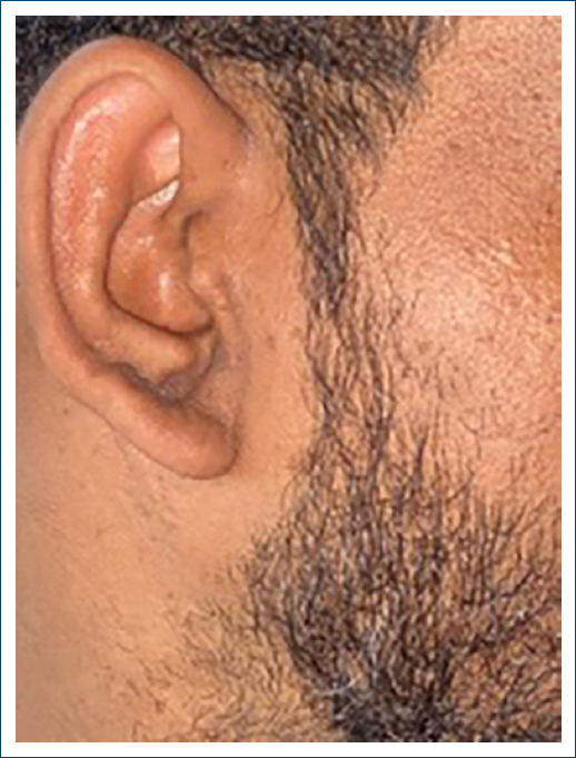
Figure 1. Thick, hyperpigmented, velvety plaque on right ear lobule.
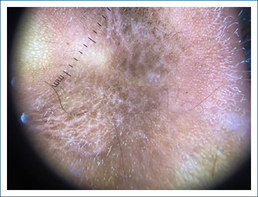
Figure 2. Polarized dermoscopy, ×200 magnification, linear crista cutis, sulcus cutis with scattered black or dark brown dots, papillary projections, and crypts.
To conclude, this case is different as the patient has acanthosis on an unusual site such as lobules of both ears and it was associated with insulin resistance, diabetes, obesity, dyslipidemia, and Vitamin D3 and Vitamin B12 deficiency. Therefore, in rare site presentation, a high index of suspicion should arise and an extensive workup is recommended as it may be associated with metabolic syndrome.
Funding
None.
Conflicts of interest
None.
Ethical considerations
Protection of human subjects and animals. The authors declare that no experiments on humans or animals were performed for this research.
Confidentiality, informed consent, and ethical approval. The authors have followed their institution’s confidentiality protocols, obtained informed consent from all patients, and secured approval from the Ethics Committee. SAGER guidelines have been followed as applicable to the nature of the study.
Declaration on the use of artificial intelligence (AI). The authors declare that no generative artificial intelligence was used in the writing or creation of the content of this manuscript.
