

Introduction
Pityriasis lichenoides et varioliformis acuta (PLEVA) is a rare dermatological condition of uncertain etiology, predominantly affecting young adults. Proposed triggers include medication use, vaccinations, and inflammatory responses to infectious agents, notably the human immunodeficiency virus (HIV).1
Clinically, PLEVA manifests as the sudden onset of multiple erythematous macules that progress into pustules or vesicles containing hemorrhagic fluid, often culminating in central ulcerations. Systemic symptoms can be present or absent alongside dermatological presentation.2,3 Diagnosis relies on clinical assessment corroborated by histopathological findings.4 However, histopathological examination may not be immediately available in all clinical settings, highlighting the importance of a noninvasive diagnostic tool. Dermoscopy is an invaluable tool in the early diagnosis of this condition, offering distinct features that aid in differential diagnosis.2,5 We present a case of HIV-associated PLEVA, confirmed through histopathology, with an exuberant presentation of lesions displaying dermoscopic features consistent with the condition.
Case report
A 27-year-old male patient with a confirmed HIV diagnosis presented with a 3-week history of sudden onset erythematous macules evolving into papules and plaques with ulceronecrotic surfaces. The lesions predominantly affected the trunk and upper limbs (Fig. 1A–C).
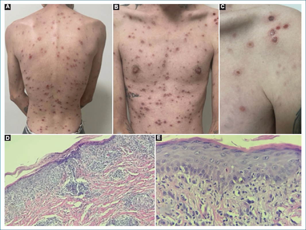
Figure 1. Clinical and histopathological features of the lesions. A-C: multiple erythematous-violaceous macules and papules, some with an ulceronecrotic surface, predominantly located on the upper limbs and trunk. Note the lesions in different stages of progression in C. D and E: histopathological findings showing a horizontal band infiltrate at the dermoepidermal junction, perivascular infiltration, parakeratosis, exocytosis of lymphocytes and neutrophils into the stratum corneum, forming corneal microabscesses.
Dermoscopy was performed using a polarized handheld dermatoscope at ×10 magnification and revealed distinct morphological patterns. The most characteristic was target-like lesions composed of a peripheral vascular ring, intermediate white scales, and a red- brown central area (Figs. 2A and B). Additional findings included lesions with a central hematic crust, surrounded by white peripheral scaling and a faint erythematous halo (Fig. 2C). The third most observed pattern consisted of lesions with minimal or absent erythematous halos, fine circular white scaling, and a vascular network of irregular linear vessels occasionally forming vascular loops (Fig. 2D).
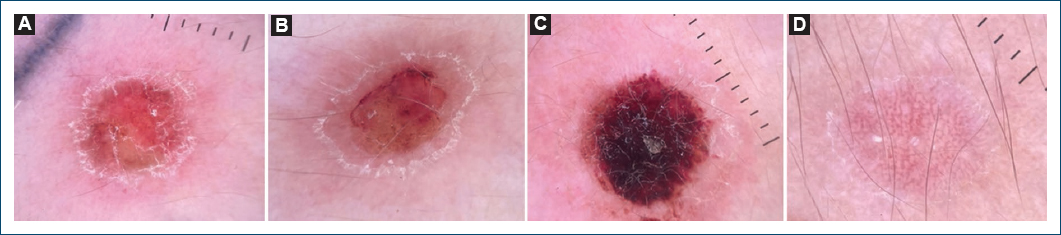
Figure 2. Dermoscopic features of the lesions. A and B: target-like pattern formed by a peripheral vascular outer ring followed by intermediate white scales and a center with a yellow-brownish mass. C: center formed by a hematinic crust, followed by a subtle peripheral white scale, surrounded by a slight erythematous halo. D: discreet erythematous vascular halo and thin circular white scale, encircling a vascular network composed of irregular linear vessels, occasionally forming vascular loops.
Histopathology confirmed the diagnosis of PLEVA, demonstrating a vacuolar interface dermatitis with features such as parakeratosis, lymphocytic and neutrophilic exocytosis, and the formation of corneal microabscesses (Figs. 1D and E).
Conclusion
Dermoscopy represents a valuable adjunctive diagnostic tool in cases of PLEVA, facilitating differentiation from clinically similar conditions such as viral exanthems, varicella, and guttate psoriasis.5 The dermoscopic findings observed in our patient are largely consistent with those previously described in the literature, including the well-established target-like pattern considered pathognomonic by several authors. In addition, we describe a dermoscopic presentation not previously reported, characterized by a central vascular pattern composed of tortuous linear vessels. These observations reinforce the relevance of dermoscopy as a rapid, noninvasive bedside tool. Further studies are warranted to better characterize dermoscopic patterns of PLEVA and to support the development of standardized diagnostic criteria.
Funding
This research has not received any specific grant from agencies in the public, commercial, or for-profit sectors.
Conflicts of interest
The authors declare no conflicts of interest.
Ethical considerations
Protection of human subjects and animals. The authors declare that no experiments on humans or animals were performed for this research.
Confidentiality, informed consent, and ethical approval. The authors have followed their institution’s confidentiality protocols, obtained informed consent from all patients, and secured approval from the Ethics Committee. SAGER guidelines have been followed as applicable to the nature of the study.
Declaration on the use of artificial intelligence. The authors declare that no generative artificial intelligence was used in the writing or creation of the content of this manuscript.
